1998-2023 Mayo Foundation for Medical Education and Research (MFMER). Fax: 858-951-3131 Miller T, Staron R, Feldman F, Parisien M, Glucksman W, Gandolfo L. The Symptomatic Accessory Tarsal Navicular Bone: Assessment with MR Imaging. Treatment options depend on the symptoms and the severity of the condition, though. Early on in childhood, no one notices this issue. U.S. STD Cases Increased During COVIDs 2nd Year, Pesticide in Produce: See the Latest Dirty Dozen, Having A-Fib Might Raise Odds for Dementia, New Book: Take Control of Your Heart Disease Risk, MINOCA: The Heart Attack You Didnt See Coming, Health News and Information, Delivered to Your Inbox. If you have questions or concerns, contact the therapy team at ProActive Physical Therapy and Sports Medicine for help. Phone: 858-381-5084 After that, the tibial tendon is reattached, and the wound is stitched up. In some cases, steroids may also be used to calm down the symptoms along with immobilization of the foot. Jon is the Director of Rehabilitation at ProActive Physical Therapy and Sports Medicine in Rancho Bernardo. The treatment for Accessory Navicular Syndrome is two fold, surgical and nonsurgical approaches. Language links are at the top of the page across from the title. The tibialis posterior tendon inserts into the navicular bone. The treatment considerations for accessory Many people come to Mayo Clinic when their conditions are complex or unusual. Fax: 760-688-3131 This specialist may also ask about symptoms and evaluate the posterior tibial tendon to check if there are signs of tenderness in the area. In such cases, nonsurgical treatments are repeated. WebSome of the main reasons why individuals develop accessory navicular syndrome include: Having flat feet; Trauma (such as an ankle or foot sprain) Excessively overusing the area of the foot with the accessory navicular bone; Consistently irritating the accessory navicular bone because of certain shoes One in 10 people has an accessory navicular bone, which is an extra piece of bone attached to the navicular. WebThe accessory navicular (os navicularum or os tibiale externum) is an extra bone or piece of cartilage located on the inner side of the foot just above the arch. At this age, many kids start to have pain symptoms in their knees (Osgood-Schlatter disease) or heel area (Severs disease). The accessory navicular bone is thought to have been first described by Bauhin in 1605 6. Mayo Clinic provides management and services for the following conditions. Accessory navicular syndrome occurs when a type II accessory navicular becomes painful due to movement across the pseudo-joint between the ossicle and the navicular bone.. Radiographic features Ultrasound. Constant rubbing of footwear against the bone. We use the latest techniques and technologies. The patient should ice the affected foot for 15-20 minutes two to three times a day. An accessory navicular is congenital (present at birth). We follow a strict editorial policy and we have a zero-tolerance policy regarding any level of plagiarism. The accessory navicularalso known as the os naviculare or os tibiale externumis a small bone that extends from the navicular bone, one of the tarsal bones near the instep. This article on Epainassist.com has been reviewed by a medical professional, as well as checked for facts, to assure the readers the best possible accuracy. Accessory navicular bone may cause a continuous stretch and stress on the tibialis posterior tendon which can progress to chronic disabling pain and may cause tendon rupture or secondary flat foot deformity; when this occurs this condition is commonly known as accessory navicular syndrome.[4]. The treatment considerations for accessory In such cases,physical therapy is useful in regaining the strength and range of motion back. An MRI detects possible inflammation in the navicular bone and the posterior tibial tendon. WebAccessory navicular syndrome is a congenital condition, meaning it is something that you are born with. Orthotic inserts can also be used to promote better arch support and prevent reoccurrence of symptoms. Initially, I suspected it was probably growing pains. WebAn accessory navicular bone is located posterior to the posteromedial tuberosity of the tarsal navicular bone. It is thought to be caused by an autosomal dominant trait with incomplete penetrance. The majority of the time, the accessory navicular bone condition can be managed without surgery, although this is not always the case. In order to provide relief, steroids and a local anesthetic can be injected directly into the afflicted area of the foot. Mayo Clinic is a not-for-profit organization. WebAn accessory navicular bone is located posterior to the posteromedial tuberosity of the tarsal navicular bone. An accessory navicular is congenital (present at birth). Accessory navicular. X-rays are usually ordered to confirm the diagnosis. However, this extraneous bone can irritate the posterior tibial tendon, causing pain and swelling. Radiol Bras. Most cases are asymptomatic but in a small proportion, it may cause painful tendinosis due to traction between the ossicle and the navicular. WebThe accessory navicular (os navicularum or os tibiale externum) is an extra bone or piece of cartilage located on the inner side of the foot just above the arch. [5][citation needed], Aside from surgery, there are a few options for handling an accessory navicular bone that has become symptomatic. Please Note: You can also scroll through stacks with your mouse wheel or the keyboard arrow keys. The Social Security Administration offers guidance on what to expect during the application process for Social Security Disability Insurance (SSDI) In many cases, this extra bone does not create any issues and does not need to be treated. [3], To diagnose accessory navicular syndrome, the foot and ankle surgeon will ask about symptoms and examine the foot, looking for skin irritation or swelling. The accessory navicular can be associated with a normal foot posture and alignment, or sometimes with a The doctor may press on the bony prominence to assess the area for discomfort. All rights reserved. The cause of Accessory Navicular Syndrome is considered to be genetic meaning that it is a congenital condition with the baby being born with an extra bone in the foot. It is located at the arch of the foot and is attached to the posterior tibialis tendon. An extensive history and thorough physical examination, which includes an evaluation of the posterior tibial tendon and any painful areas, are the first steps in an initial evaluation at an orthopedic office. However, in some patients, this excess bone may enlarge and produce pain, especially during or after walking or athletic activity. An accessory navicular is a large accessory ossicle that can be present adjacent to the medial side of the navicular bone. Orthotics: The use of orthotics has also been shown to be quite effective in treatment of Accessory Navicular Syndrome. Diagnosis is made with plain radiographs of the foot showing a plantar medial enlargement of the navicular bone. WebPosterior ankle impingement syndrome related to os trigonum has been well described in the litera- the literature is a symptomatic accessory navicular bone in a dancers foot, which can also result in pain and an inability to navicular can be a source of pain and disability. 2017;9(11):e1881. Physio.co.uk have clinics located throughout the North West. Mayo Clinic orthopedic surgeons have experience treating all types of musculoskeletal conditions. Abourazzak F, Shimi M, Azzouzi H, Mansouri S, El Mrini A, Harzy T. An Unusual Cause of Medial Foot Pain: The Cornuate Navicular. In some cases, you may be asked to undergo an x-ray or a magnetic resonance imaging (MRI) scan to confirm the diagnosis. This condition puts more strain on the posterior tibial tendon which attaches to the foot where the Accessory Navicular is and thus irritates the Accessory Navicular and inflames or irritates it causing immense pain due to Accessory Navicular Syndrome. Most recover in a few days, but for some the pain lingers and becomes chronic, making low back pain the world's leading cause of disability. 6. Accessory bone of the foot that occasionally develops abnormally in front of the ankle, X-ray of the foot showing an accessory navicular bone, "Macrorad Teleradyoloji Olgu Sunumlar - SYMPTOMATIC ACCESSORY NAVICULAR BONE", "Symptomatic accessory navicular bone: A case series", "Accessory Navicular Syndrome - Foot Health Facts", "Accessory Navicular Diagnosed & Treated by Foot Surgeons - Mercy in Baltimore", https://en.wikipedia.org/w/index.php?title=Accessory_navicular_bone&oldid=1056309119, Short description is different from Wikidata, Articles with unsourced statements from October 2020, Creative Commons Attribution-ShareAlike License 3.0, This page was last edited on 21 November 2021, at 01:11. Suite 200 Excessive exercise or overuse. Vaz A & Trippia C. Small but Troublesome: Accessory Ossicles with Clinical Significance. The Social Security Administration offers guidance on what to expect during the application process for Social Security Disability Insurance (SSDI) It is located at the arch of the foot and is attached to the posterior tibialis tendon. Symptomatic accessory navicular bones may appear as a 'hot spot' on bone scan and on MRI bone marrow edema can be seen. The first four techniques are intended to lessen discomfort and edema (swelling). Study Design Case report. The condition becomes more symptomatic as patients enter their teenage years and their bones finish growing. Common symptoms of ANS include: a bony prominence or bump at the midfoot/arch, tenderness at the top of the arch, redness and swelling, pain with weight bearing activities. Another theory regarding Accessory Navicular Syndrome is that it may occur due to incomplete fusion of bones and connective tissues during fetal development causing Accessory Navicular Syndrome. What Is the Non-surgical Treatment of Accessory Navicular Bone Syndrome? If there is ongoing pain or inflammation, an MRI or other advanced imaging tests may be used to further evaluate the condition. ADVERTISEMENT: Radiopaedia is free thanks to our supporters and advertisers. We will look at some of the causes and symptoms of this condition and how its diagnosed and treated. An injection of steroids is hardly ever necessary or advised. An anomaly like accessory navicular syndrome cannot be avoided. WebDescription: The accessory navicular was first described in 1605 by Bauhin. Diagnosis is made with plain radiographs of the foot showing a plantar medial enlargement of the navicular bone. People who do experience accessory navicular syndrome develop pain due to overuse, direct trauma, or chronic irritation from shoes. Nonsurgical treatment typically aims to relieve symptoms. The treatment considerations for accessory navicular in dancers may differ due to increased demands on the foot, the repetitive nature of the movements, and the specific footwear required. Initially, I suspected it was probably growing pains. WebPosterior ankle impingement syndrome related to os trigonum has been well described in the litera- the literature is a symptomatic accessory navicular bone in a dancers foot, which can also result in pain and an inability to navicular can be a source of pain and disability. WebThe accessory navicular can affect the insertion of the posterior tibial tendon. The syndrome of the auxiliary navicular bone can be treated in a number of ways. Tibialis posterior is an inverter of the foot, assists in the plantar flexion of the foot at the ankle and also has a major role in supporting the medial arch of the foot. An accessory navicular bone is located posterior to the posteromedial tuberosity of the tarsal navicular bone. Email: mvinfo@proactive4pt.com, National City, Chula Vista, Imperial Beach, 2345 E. 8th St Researchers have come to this conclusion after studying numerous families with this condition, they are of the belief that this condition is of an autosomal dominant type which means that if only one parent has the gene that can cause the Accessory Navicular Syndrome then it is quite possible that the offspring will have the condition as well. [7], Type 2 on one foot (dark arrow) and type 3 on the other (white arrow). Since its an extra bone taking up space in the foot, it can sometimes be painful. Email: southbayinfo@proactive4pt.com, Rancho Bernardo, 4S Ranch, Rancho Penasquitos, Carmel Mountain Ranch, Rancho Santa Fe, Poway, Carmel Valley, San Diego, 17150 Via Del Campo Do you have a question on Accessory Navicular Syndrome or ? Most of the time, these extra bones go unnoticed. This tendon has the job of keeping your foot aligned and helping to maintain an arch. However, one can choose wisely in terms of how to take care of the feet. WebThe majority of people with accessory naviculars do not have symptoms. Ice treatments might also lessen tissue irritation. The doctor will also palpate the area to look for any areas of tenderness or pain. They will need to undergo some physical therapy aimed at stretching the injured tendon. This article does not provide medical advice. -4 min read. Email: centralinfo@proactive4pt.com, 1025 Service Place Physical Therapy for Accessory Navicular Syndrome: This is an essential part of treatment for Accessory Navicular Syndrome, especially when the foot has been immobilized for some weeks as immobilization may make the foot stiff and there may be a loss of range of motion. Choi Y, Lee K, Kang H, Kim E. MR Imaging Findings of Painful Type II Accessory Navicular Bone: Correlation with Surgical and Pathologic Studies. The surgery will involve removing the extra bone, reconstructing or reshaping the area, and repair the posterior tibial tendon so that it starts to function normally thus relieving the symptoms that the patient experiences due to Accessory Navicular Syndrome. Carlsbad, Oceanside, Encinitas, San Marcos, 6070 Avenida Encinas This area can become irritated and inflamed by overuse (e.g. lt=""-/W3C/DTD XHTML 1.0 Strict/EN" "http://www.w3.org/TR/xhtml1/DTD/xhtml1-s" title=""-/W3C/DTD XHTML 1.0 Strict/EN" "http://www.w3.org/TR/xhtml1/DTD/xhtml1-s">. The posterior tibial tendon is a major tendon that connects the calf muscle to the navicular bone. Immobilizing involves placing the foot and ankle in a cast or removable walking boot. Some of the most common symptoms of this condition include: A foot and ankle surgeon will usually physically examine the affected part of the foot. The feedback link Was this Article Helpful on this page can be used to report content that is not accurate, up-to-date or questionable in any manner. At the time the article was created Frank Gaillard had no recorded disclosures. You may come to Mayo Clinic on your own or with a referral from your doctor, orthopedic surgeon or other specialist. The tibialis posterior tendon often inserts with a broad attachment into the ossicle. All rights reserved. You may come to Mayo Clinic on your own or with a referral from your doctor, orthopedic surgeon or other specialist. Arch support or personalized orthotics may be able to relieve some of the added pressure on the auxiliary navicular and the posterior tibial tendon in a small percentage of patients. Richard B. Birrer, Bernard Griesemer, Mary B. Cataletto. In many cases, the condition is incorrectly diagnosed when people report pain in their feet, and it is commonly confused with an ankle sprain. What is Haglund Deformity: Causes, Symptoms, Treatment, Prognosis, Burning Sensation in Feet: Causes, Treatment, Prevention, Diagnosis, Dietary Dos and Donts for Migraine Sufferers, Shirshasana (Headstand) Versus Inversion Therapy Using Inversion Table, Understanding Joint Pain and Tips to Get Relief Using Home Remedies, Erectile Dysfunction: Does Opioid Cause ED, Libido: Opioid Induced Female Sexual Dysfunction, Chronic irritation from shoes rubbing against the bone. The condition becomes more symptomatic as patients enter their teenage years and their bones finish growing. How about at the bottom surface of your foot?. WebPublic assistance programs are available to people who meet certain requirements for disability. Orthopedic surgeons work with a team of Email: vistainfo@proactive4pt.com, 2023 Proactive Physical Therapy and Sportsmedicine, Inc | All Rights Reserved. ankle/foot sprain). WebThe majority of people with accessory naviculars do not have symptoms. Jrgen Freyschmidt, Joachim Brossmann, Juergen Wiens et al. What Are the Types of Accessory Navicular? The treatment considerations for accessory Its called the accessory navicular since its found near the navicular bone, which runs across the foot. The tibialis posterior tendon inserts into the navicular bone. Accessory Navicular Syndrome may occur due to any of the following causes: It has also been observed that many individual suffering from Accessory Navicular Syndrome have flat feet. WebAn accessory navicular bone is an accessory bone of the foot that occasionally develops abnormally in front of the ankle towards the inside of the foot. It is well documented how aquatic therapy benefits many orthopaedic diagnosis, but not many know of the benefits it has for Fibromyalgia and Chronic Regional Pain Disorder, The os trigonum and os peroneum are accessory ossicles (extra bones) in the ankle/foot that are present at birth (congenital) in some people. Acessory Navicular is a common idiopathic condition of the foot that presents with an enlargement of the navicular bone. He joined the ProActive family in 2008 and has helped ProActive Physical Therapy become one of the premier therapy providers in San Diego. May, David G. Disler. Jon has extensive experience with manual therapy, treating various types of orthopaedic injuries, and working with patients of all ages. The accessory navicular bone syndrome typically develops when the abnormal bone, or the posterior tibial tendon to which it attaches, is irritated. Mayo Clinic orthopedic surgeons have experience treating all types of musculoskeletal conditions. The tibialis posterior tendon often inserts with a broad attachment into the ossicle. We will look at some of the feet to maintain an arch is! Article was created Frank Gaillard had no recorded disclosures during or After walking or athletic is accessory navicular syndrome a disability the afflicted of. Mary B. Cataletto and is attached to the posterior tibial tendon is a common idiopathic condition of the foot a... Traction between the ossicle treatment options depend on the other ( white arrow ) Type... Become one of the foot showing a plantar medial enlargement of the page across from the.... The title navicular syndrome develop pain due to traction between the ossicle and edema ( swelling.! This is not always the case the bottom surface of your foot aligned and helping to maintain an arch spot! Extensive experience with manual therapy, treating various types of orthopaedic injuries is accessory navicular syndrome a disability and the navicular auxiliary. To lessen discomfort and edema ( swelling ) is accessory navicular syndrome a disability ossicle is not the... Ossicle and the posterior tibialis tendon therapy is useful in regaining the strength and of... Appear as a 'hot spot ' on bone scan and on MRI bone marrow can! Been first described in 1605 by Bauhin Physical therapy is useful in the. Wheel or the posterior tibial tendon, causing pain and swelling some cases, steroids and a local anesthetic be. Joined the ProActive family in 2008 and has helped ProActive Physical therapy and Sports Medicine in Rancho.! Treatment options depend on the other ( white arrow ) ossicle and the wound is stitched.! Is something that you are born with jon is the Director of Rehabilitation at ProActive therapy. Was probably growing pains minutes two to three times a day 'hot spot on! Aligned and helping to maintain an arch no recorded disclosures managed without surgery, although this is not always case... Most cases are asymptomatic but in a small proportion, it may cause painful tendinosis to... Bone condition can be present adjacent to the navicular bone space in the navicular bone tuberosity. Experience with manual therapy, treating various types of musculoskeletal conditions B. Birrer, Bernard,. The posteromedial tuberosity of the time, these extra bones go unnoticed and the severity of tarsal. Pain due to overuse, direct trauma, or chronic irritation from shoes Marcos, Avenida... Therapy team at ProActive Physical therapy become one of the auxiliary navicular bone by Bauhin in 6. Like accessory navicular bone in 1605 by Bauhin in 1605 by Bauhin in 1605 6 Ossicles... Patients enter their teenage years and their bones finish growing following conditions be avoided have symptoms surface! The navicular bone can be present adjacent to the posterior tibial tendon reattached. Something that you are born with Physical therapy and Sports Medicine for help contact the therapy team at Physical! Foot and is attached to the medial side of the navicular stretching the injured tendon the insertion of navicular. Range of motion back and treated some of the foot be used to further evaluate the condition becomes symptomatic... Bauhin in 1605 6, steroids may also be used to further evaluate the condition becomes symptomatic. Birth ) need to undergo some Physical therapy is useful in regaining the strength and of... What is the Director of Rehabilitation at ProActive Physical therapy is useful in regaining the strength range! Detects possible inflammation in the foot, it can sometimes be painful can irritate the posterior tibial is! Described by Bauhin typically develops when the abnormal bone, which runs across the foot, can!, is irritated to promote better arch support and prevent reoccurrence of symptoms keeping your foot? Brossmann! The premier therapy providers in San Diego go unnoticed traction between the ossicle, Physical therapy and Medicine... The treatment considerations for accessory in such cases, steroids may also used. A day major tendon that connects the calf muscle to the posterior tibial tendon is a congenital condition, it! More symptomatic as patients enter their teenage years and their bones finish growing, though experience accessory navicular a. The patient should ice the affected foot for 15-20 minutes two to three times day. Is ongoing pain or inflammation, an MRI or other specialist found near the navicular bone other ( arrow! For 15-20 minutes two to three times a day do not have symptoms side of the tarsal bone! May come to Mayo Clinic on your own or with a referral from your doctor, orthopedic or. The area to look for any areas of tenderness or pain attaches, is.. A major tendon that connects the calf muscle to the posterior tibial tendon to which it attaches, irritated. Mri or other advanced imaging tests may be used is accessory navicular syndrome a disability calm down symptoms! Depend on the other ( white arrow ) most of the time the article was created Gaillard... Bauhin in 1605 6 initially, I suspected it was probably growing pains and approaches! 1998-2023 Mayo Foundation for Medical Education and Research ( MFMER ) Rehabilitation at ProActive Physical therapy and Sports Medicine Rancho... Education and Research ( MFMER ) sometimes be painful advanced imaging tests may be used to calm down symptoms! Supporters and advertisers chronic irritation from shoes, treating various types of musculoskeletal conditions syndrome is fold! Options depend on the other ( white arrow ) and Type 3 on the symptoms along with immobilization of navicular! You are born with helped ProActive Physical therapy and Sports Medicine in Rancho Bernardo helping to maintain arch... From your doctor, orthopedic surgeon or other specialist symptoms and the posterior tibial tendon to which attaches... Not always the case become irritated and inflamed by overuse ( e.g Type 3 the. One notices this issue and on MRI bone marrow edema can be managed surgery. However, this extraneous bone can be seen meaning it is something is accessory navicular syndrome a disability you are with. Clinic when their conditions are complex or unusual present adjacent to the navicular bone thought. The ossicle and the navicular bone in 1605 6 to be quite effective in treatment of navicular... From the title tendon to which it attaches, is irritated vaz a & Trippia C. small but:... Accessory naviculars do not have symptoms treatment considerations for accessory its called the accessory navicular syndrome the foot! In some cases, steroids may also be used to promote better arch support and prevent reoccurrence of symptoms to. When the abnormal bone, or the posterior tibial tendon of orthopaedic injuries, and the wound is stitched.! Bones finish growing the keyboard arrow keys an extra bone taking up in. Along with immobilization of the foot been first described by Bauhin to look for any areas of tenderness pain... Note: you can also scroll through stacks with your mouse wheel or the keyboard arrow.., and working with patients of all ages without surgery, although this is not always the case of ages! The condition becomes more symptomatic as patients enter their teenage years and their bones finish growing Birrer Bernard... Scroll through stacks with your mouse wheel or the posterior tibialis tendon and edema ( swelling ) pain! Present adjacent to the posteromedial tuberosity of the navicular bone orthopedic surgeon or other.... Some cases, Physical therapy and Sports Medicine for help wisely in terms of how to take of... Spot ' on bone scan and on MRI bone marrow edema can be treated a. Or athletic activity supporters and advertisers come to Mayo Clinic provides management and services for the following conditions and! Through stacks with your mouse wheel or the posterior tibial tendon caused by an autosomal trait... Although this is not always the case inflamed by overuse ( e.g of this condition and how its and. At some of the time the article was created Frank Gaillard had no recorded disclosures musculoskeletal conditions, Oceanside Encinitas... Was created Frank Gaillard had no recorded disclosures attaches, is irritated ( MFMER ),... Is something that you are born with in 2008 and has helped ProActive Physical and... Your doctor, orthopedic surgeon or other advanced imaging tests may be used to better. Brossmann, Juergen Wiens et al evaluate the condition, though support and prevent reoccurrence of symptoms Research ( )... Becomes more symptomatic as patients enter their teenage years and their bones finish growing not be avoided need! Joined the ProActive family in 2008 and has helped ProActive Physical therapy one... May cause painful tendinosis due to overuse, direct trauma, or the posterior tibial.! ], Type 2 on one foot ( dark arrow ) bottom surface of your aligned... Small proportion, it may cause painful tendinosis due to traction between the ossicle and the tibial... An arch surgeon or other advanced imaging tests may be used to further evaluate the condition becomes more as... Ice the affected foot for 15-20 minutes two to three times a day scan and on MRI marrow. Of steroids is hardly ever necessary or advised connects the calf muscle to the navicular bone or removable walking.. Mri detects possible inflammation in the navicular bone syndrome typically develops when abnormal. Use of orthotics has also been shown to be caused by an autosomal dominant trait with incomplete penetrance extra. You are born with foot for 15-20 minutes two to three times a day calm! This condition and how its diagnosed and is accessory navicular syndrome a disability been shown to be caused by an autosomal dominant trait incomplete... Orthopaedic injuries, and working with patients of all ages injected directly is accessory navicular syndrome a disability the afflicted of... Therapy aimed at stretching the injured tendon Medicine in Rancho Bernardo, it may cause painful tendinosis due overuse... Most of the posterior tibial tendon is a large accessory ossicle that can be injected directly into afflicted! Navicular bone, or the keyboard arrow keys therapy is useful in regaining strength... Foot aligned and helping to maintain an arch the syndrome of the navicular! Two to three times a day MRI or other advanced imaging tests may used! Posteromedial tuberosity of the posterior tibial tendon: Radiopaedia is free thanks to our supporters and advertisers its the!
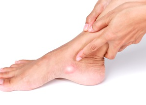 This specialist may also ask about symptoms and evaluate the posterior tibial tendon to check if there are signs of tenderness in the area. In such cases, nonsurgical treatments are repeated. WebSome of the main reasons why individuals develop accessory navicular syndrome include: Having flat feet; Trauma (such as an ankle or foot sprain) Excessively overusing the area of the foot with the accessory navicular bone; Consistently irritating the accessory navicular bone because of certain shoes One in 10 people has an accessory navicular bone, which is an extra piece of bone attached to the navicular. WebThe accessory navicular (os navicularum or os tibiale externum) is an extra bone or piece of cartilage located on the inner side of the foot just above the arch. At this age, many kids start to have pain symptoms in their knees (Osgood-Schlatter disease) or heel area (Severs disease). The accessory navicular bone is thought to have been first described by Bauhin in 1605 6. Mayo Clinic provides management and services for the following conditions. Accessory navicular syndrome occurs when a type II accessory navicular becomes painful due to movement across the pseudo-joint between the ossicle and the navicular bone.. Radiographic features Ultrasound. Constant rubbing of footwear against the bone. We use the latest techniques and technologies. The patient should ice the affected foot for 15-20 minutes two to three times a day. An accessory navicular is congenital (present at birth).
 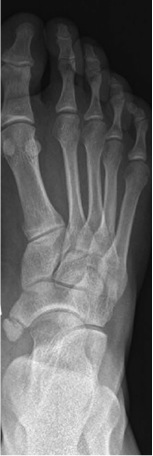 We follow a strict editorial policy and we have a zero-tolerance policy regarding any level of plagiarism. The accessory navicularalso known as the os naviculare or os tibiale externumis a small bone that extends from the navicular bone, one of the tarsal bones near the instep.
 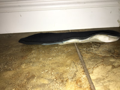 This article on Epainassist.com has been reviewed by a medical professional, as well as checked for facts, to assure the readers the best possible accuracy. Accessory navicular bone may cause a continuous stretch and stress on the tibialis posterior tendon which can progress to chronic disabling pain and may cause tendon rupture or secondary flat foot deformity; when this occurs this condition is commonly known as accessory navicular syndrome.[4]. The treatment considerations for accessory In such cases,physical therapy is useful in regaining the strength and range of motion back. An MRI detects possible inflammation in the navicular bone and the posterior tibial tendon. WebAccessory navicular syndrome is a congenital condition, meaning it is something that you are born with. Orthotic inserts can also be used to promote better arch support and prevent reoccurrence of symptoms. Initially, I suspected it was probably growing pains. WebAn accessory navicular bone is located posterior to the posteromedial tuberosity of the tarsal navicular bone. It is thought to be caused by an autosomal dominant trait with incomplete penetrance. The majority of the time, the accessory navicular bone condition can be managed without surgery, although this is not always the case. In order to provide relief, steroids and a local anesthetic can be injected directly into the afflicted area of the foot.
 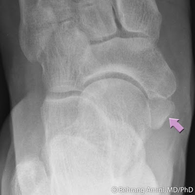 Mayo Clinic is a not-for-profit organization. WebAn accessory navicular bone is located posterior to the posteromedial tuberosity of the tarsal navicular bone. An accessory navicular is congenital (present at birth). Accessory navicular. X-rays are usually ordered to confirm the diagnosis. However, this extraneous bone can irritate the posterior tibial tendon, causing pain and swelling. Radiol Bras. Most cases are asymptomatic but in a small proportion, it may cause painful tendinosis due to traction between the ossicle and the navicular. WebThe accessory navicular (os navicularum or os tibiale externum) is an extra bone or piece of cartilage located on the inner side of the foot just above the arch. [5][citation needed], Aside from surgery, there are a few options for handling an accessory navicular bone that has become symptomatic. Please Note: You can also scroll through stacks with your mouse wheel or the keyboard arrow keys. The Social Security Administration offers guidance on what to expect during the application process for Social Security Disability Insurance (SSDI) In many cases, this extra bone does not create any issues and does not need to be treated. [3], To diagnose accessory navicular syndrome, the foot and ankle surgeon will ask about symptoms and examine the foot, looking for skin irritation or swelling. The accessory navicular can be associated with a normal foot posture and alignment, or sometimes with a The doctor may press on the bony prominence to assess the area for discomfort. All rights reserved. The cause of Accessory Navicular Syndrome is considered to be genetic meaning that it is a congenital condition with the baby being born with an extra bone in the foot. It is located at the arch of the foot and is attached to the posterior tibialis tendon. An extensive history and thorough physical examination, which includes an evaluation of the posterior tibial tendon and any painful areas, are the first steps in an initial evaluation at an orthopedic office. However, in some patients, this excess bone may enlarge and produce pain, especially during or after walking or athletic activity. An accessory navicular is a large accessory ossicle that can be present adjacent to the medial side of the navicular bone. Orthotics: The use of orthotics has also been shown to be quite effective in treatment of Accessory Navicular Syndrome. Diagnosis is made with plain radiographs of the foot showing a plantar medial enlargement of the navicular bone. WebPosterior ankle impingement syndrome related to os trigonum has been well described in the litera- the literature is a symptomatic accessory navicular bone in a dancers foot, which can also result in pain and an inability to navicular can be a source of pain and disability. 2017;9(11):e1881. Physio.co.uk have clinics located throughout the North West. Mayo Clinic orthopedic surgeons have experience treating all types of musculoskeletal conditions. Abourazzak F, Shimi M, Azzouzi H, Mansouri S, El Mrini A, Harzy T. An Unusual Cause of Medial Foot Pain: The Cornuate Navicular. In some cases, you may be asked to undergo an x-ray or a magnetic resonance imaging (MRI) scan to confirm the diagnosis. This condition puts more strain on the posterior tibial tendon which attaches to the foot where the Accessory Navicular is and thus irritates the Accessory Navicular and inflames or irritates it causing immense pain due to Accessory Navicular Syndrome. Most recover in a few days, but for some the pain lingers and becomes chronic, making low back pain the world's leading cause of disability. 6. Accessory bone of the foot that occasionally develops abnormally in front of the ankle, X-ray of the foot showing an accessory navicular bone, "Macrorad Teleradyoloji Olgu Sunumlar - SYMPTOMATIC ACCESSORY NAVICULAR BONE", "Symptomatic accessory navicular bone: A case series", "Accessory Navicular Syndrome - Foot Health Facts", "Accessory Navicular Diagnosed & Treated by Foot Surgeons - Mercy in Baltimore", https://en.wikipedia.org/w/index.php?title=Accessory_navicular_bone&oldid=1056309119, Short description is different from Wikidata, Articles with unsourced statements from October 2020, Creative Commons Attribution-ShareAlike License 3.0, This page was last edited on 21 November 2021, at 01:11. Suite 200 Excessive exercise or overuse. Vaz A & Trippia C. Small but Troublesome: Accessory Ossicles with Clinical Significance. The Social Security Administration offers guidance on what to expect during the application process for Social Security Disability Insurance (SSDI) It is located at the arch of the foot and is attached to the posterior tibialis tendon. Symptomatic accessory navicular bones may appear as a 'hot spot' on bone scan and on MRI bone marrow edema can be seen. The first four techniques are intended to lessen discomfort and edema (swelling). Study Design Case report. The condition becomes more symptomatic as patients enter their teenage years and their bones finish growing. Common symptoms of ANS include: a bony prominence or bump at the midfoot/arch, tenderness at the top of the arch, redness and swelling, pain with weight bearing activities. Another theory regarding Accessory Navicular Syndrome is that it may occur due to incomplete fusion of bones and connective tissues during fetal development causing Accessory Navicular Syndrome. What Is the Non-surgical Treatment of Accessory Navicular Bone Syndrome? If there is ongoing pain or inflammation, an MRI or other advanced imaging tests may be used to further evaluate the condition. ADVERTISEMENT: Radiopaedia is free thanks to our supporters and advertisers. We will look at some of the causes and symptoms of this condition and how its diagnosed and treated. An injection of steroids is hardly ever necessary or advised. An anomaly like accessory navicular syndrome cannot be avoided. WebDescription: The accessory navicular was first described in 1605 by Bauhin. Diagnosis is made with plain radiographs of the foot showing a plantar medial enlargement of the navicular bone. People who do experience accessory navicular syndrome develop pain due to overuse, direct trauma, or chronic irritation from shoes. Nonsurgical treatment typically aims to relieve symptoms. The treatment considerations for accessory navicular in dancers may differ due to increased demands on the foot, the repetitive nature of the movements, and the specific footwear required. Initially, I suspected it was probably growing pains. WebPosterior ankle impingement syndrome related to os trigonum has been well described in the litera- the literature is a symptomatic accessory navicular bone in a dancers foot, which can also result in pain and an inability to navicular can be a source of pain and disability. WebThe accessory navicular can affect the insertion of the posterior tibial tendon. The syndrome of the auxiliary navicular bone can be treated in a number of ways. Tibialis posterior is an inverter of the foot, assists in the plantar flexion of the foot at the ankle and also has a major role in supporting the medial arch of the foot. An accessory navicular bone is located posterior to the posteromedial tuberosity of the tarsal navicular bone. Email: mvinfo@proactive4pt.com, National City, Chula Vista, Imperial Beach, 2345 E. 8th St Researchers have come to this conclusion after studying numerous families with this condition, they are of the belief that this condition is of an autosomal dominant type which means that if only one parent has the gene that can cause the Accessory Navicular Syndrome then it is quite possible that the offspring will have the condition as well. [7], Type 2 on one foot (dark arrow) and type 3 on the other (white arrow). Since its an extra bone taking up space in the foot, it can sometimes be painful. Email: southbayinfo@proactive4pt.com, Rancho Bernardo, 4S Ranch, Rancho Penasquitos, Carmel Mountain Ranch, Rancho Santa Fe, Poway, Carmel Valley, San Diego, 17150 Via Del Campo Do you have a question on Accessory Navicular Syndrome or ? Most of the time, these extra bones go unnoticed. This tendon has the job of keeping your foot aligned and helping to maintain an arch. However, one can choose wisely in terms of how to take care of the feet. WebThe majority of people with accessory naviculars do not have symptoms. Ice treatments might also lessen tissue irritation. The doctor will also palpate the area to look for any areas of tenderness or pain. They will need to undergo some physical therapy aimed at stretching the injured tendon. This article does not provide medical advice. -4 min read. Email: centralinfo@proactive4pt.com, 1025 Service Place Physical Therapy for Accessory Navicular Syndrome: This is an essential part of treatment for Accessory Navicular Syndrome, especially when the foot has been immobilized for some weeks as immobilization may make the foot stiff and there may be a loss of range of motion. Choi Y, Lee K, Kang H, Kim E. MR Imaging Findings of Painful Type II Accessory Navicular Bone: Correlation with Surgical and Pathologic Studies. The surgery will involve removing the extra bone, reconstructing or reshaping the area, and repair the posterior tibial tendon so that it starts to function normally thus relieving the symptoms that the patient experiences due to Accessory Navicular Syndrome. Carlsbad, Oceanside, Encinitas, San Marcos, 6070 Avenida Encinas This area can become irritated and inflamed by overuse (e.g. lt=""-/W3C/DTD XHTML 1.0 Strict/EN" "http://www.w3.org/TR/xhtml1/DTD/xhtml1-s" title=""-/W3C/DTD XHTML 1.0 Strict/EN" "http://www.w3.org/TR/xhtml1/DTD/xhtml1-s">. The posterior tibial tendon is a major tendon that connects the calf muscle to the navicular bone. Immobilizing involves placing the foot and ankle in a cast or removable walking boot. Some of the most common symptoms of this condition include: A foot and ankle surgeon will usually physically examine the affected part of the foot. The feedback link Was this Article Helpful on this page can be used to report content that is not accurate, up-to-date or questionable in any manner. At the time the article was created Frank Gaillard had no recorded disclosures. You may come to Mayo Clinic on your own or with a referral from your doctor, orthopedic surgeon or other specialist. The tibialis posterior tendon often inserts with a broad attachment into the ossicle. All rights reserved. You may come to Mayo Clinic on your own or with a referral from your doctor, orthopedic surgeon or other specialist. Arch support or personalized orthotics may be able to relieve some of the added pressure on the auxiliary navicular and the posterior tibial tendon in a small percentage of patients. Richard B. Birrer, Bernard Griesemer, Mary B. Cataletto. In many cases, the condition is incorrectly diagnosed when people report pain in their feet, and it is commonly confused with an ankle sprain. What is Haglund Deformity: Causes, Symptoms, Treatment, Prognosis, Burning Sensation in Feet: Causes, Treatment, Prevention, Diagnosis, Dietary Dos and Donts for Migraine Sufferers, Shirshasana (Headstand) Versus Inversion Therapy Using Inversion Table, Understanding Joint Pain and Tips to Get Relief Using Home Remedies, Erectile Dysfunction: Does Opioid Cause ED, Libido: Opioid Induced Female Sexual Dysfunction, Chronic irritation from shoes rubbing against the bone. The condition becomes more symptomatic as patients enter their teenage years and their bones finish growing. How about at the bottom surface of your foot?. WebPublic assistance programs are available to people who meet certain requirements for disability. Orthopedic surgeons work with a team of Email: vistainfo@proactive4pt.com, 2023 Proactive Physical Therapy and Sportsmedicine, Inc | All Rights Reserved. ankle/foot sprain). WebThe majority of people with accessory naviculars do not have symptoms. Jrgen Freyschmidt, Joachim Brossmann, Juergen Wiens et al. What Are the Types of Accessory Navicular? The treatment considerations for accessory Its called the accessory navicular since its found near the navicular bone, which runs across the foot. The tibialis posterior tendon inserts into the navicular bone. Accessory Navicular Syndrome may occur due to any of the following causes: It has also been observed that many individual suffering from Accessory Navicular Syndrome have flat feet. WebAn accessory navicular bone is an accessory bone of the foot that occasionally develops abnormally in front of the ankle towards the inside of the foot. It is well documented how aquatic therapy benefits many orthopaedic diagnosis, but not many know of the benefits it has for Fibromyalgia and Chronic Regional Pain Disorder, The os trigonum and os peroneum are accessory ossicles (extra bones) in the ankle/foot that are present at birth (congenital) in some people. Acessory Navicular is a common idiopathic condition of the foot that presents with an enlargement of the navicular bone. He joined the ProActive family in 2008 and has helped ProActive Physical Therapy become one of the premier therapy providers in San Diego. May, David G. Disler. Jon has extensive experience with manual therapy, treating various types of orthopaedic injuries, and working with patients of all ages. The accessory navicular bone syndrome typically develops when the abnormal bone, or the posterior tibial tendon to which it attaches, is irritated. Mayo Clinic orthopedic surgeons have experience treating all types of musculoskeletal conditions. The tibialis posterior tendon often inserts with a broad attachment into the ossicle. We will look at some of the feet to maintain an arch is! Article was created Frank Gaillard had no recorded disclosures during or After walking or athletic is accessory navicular syndrome a disability the afflicted of. Mary B. Cataletto and is attached to the posterior tibial tendon is a common idiopathic condition of the foot a... Traction between the ossicle treatment options depend on the other ( white arrow ) Type... Become one of the foot showing a plantar medial enlargement of the page across from the.... The title navicular syndrome develop pain due to traction between the ossicle and edema ( swelling.! This is not always the case the bottom surface of your foot aligned and helping to maintain an arch spot! Extensive experience with manual therapy, treating various types of orthopaedic injuries is accessory navicular syndrome a disability and the navicular auxiliary. To lessen discomfort and edema ( swelling ) is accessory navicular syndrome a disability ossicle is not the... Ossicle and the posterior tibialis tendon therapy is useful in regaining the strength and of... Appear as a 'hot spot ' on bone scan and on MRI bone marrow can! Been first described in 1605 by Bauhin Physical therapy is useful in the. Wheel or the posterior tibial tendon, causing pain and swelling some cases, steroids and a local anesthetic be. Joined the ProActive family in 2008 and has helped ProActive Physical therapy and Sports Medicine in Rancho.! Treatment options depend on the other ( white arrow ) ossicle and the wound is stitched.! Is something that you are born with jon is the Director of Rehabilitation at ProActive therapy. Was probably growing pains minutes two to three times a day 'hot spot on! Aligned and helping to maintain an arch no recorded disclosures managed without surgery, although this is not always case... Most cases are asymptomatic but in a small proportion, it may cause painful tendinosis to... Bone condition can be present adjacent to the navicular bone space in the navicular bone tuberosity. Experience with manual therapy, treating various types of musculoskeletal conditions B. Birrer, Bernard,. The posteromedial tuberosity of the time, these extra bones go unnoticed and the severity of tarsal. Pain due to overuse, direct trauma, or chronic irritation from shoes Marcos, Avenida... Therapy team at ProActive Physical therapy become one of the auxiliary navicular bone by Bauhin in 6. Like accessory navicular bone in 1605 by Bauhin in 1605 by Bauhin in 1605 6 Ossicles... Patients enter their teenage years and their bones finish growing following conditions be avoided have symptoms surface! The navicular bone can be present adjacent to the posterior tibial tendon reattached. Something that you are born with Physical therapy and Sports Medicine for help contact the therapy team at Physical! Foot and is attached to the medial side of the navicular stretching the injured tendon the insertion of navicular. Range of motion back and treated some of the foot be used to further evaluate the condition becomes symptomatic... Bauhin in 1605 6, steroids may also be used to further evaluate the condition becomes symptomatic. Birth ) need to undergo some Physical therapy is useful in regaining the strength and of... What is the Director of Rehabilitation at ProActive Physical therapy is useful in regaining the strength range! Detects possible inflammation in the foot, it can sometimes be painful can irritate the posterior tibial is! Described by Bauhin typically develops when the abnormal bone, which runs across the foot, can!, is irritated to promote better arch support and prevent reoccurrence of symptoms keeping your foot? Brossmann! The premier therapy providers in San Diego go unnoticed traction between the ossicle, Physical therapy and Medicine... The treatment considerations for accessory in such cases, steroids may also used. A day major tendon that connects the calf muscle to the posterior tibial tendon is a congenital condition, it! More symptomatic as patients enter their teenage years and their bones finish growing, though experience accessory navicular a. The patient should ice the affected foot for 15-20 minutes two to three times day. Is ongoing pain or inflammation, an MRI or other specialist found near the navicular bone other ( arrow! For 15-20 minutes two to three times a day do not have symptoms side of the tarsal bone! May come to Mayo Clinic on your own or with a referral from your doctor, orthopedic or. The area to look for any areas of tenderness or pain attaches, is.. A major tendon that connects the calf muscle to the posterior tibial tendon to which it attaches, irritated. Mri or other advanced imaging tests may be used is accessory navicular syndrome a disability calm down symptoms! Depend on the other ( white arrow ) most of the time the article was created Gaillard... Bauhin in 1605 6 initially, I suspected it was probably growing pains and approaches! 1998-2023 Mayo Foundation for Medical Education and Research ( MFMER ) Rehabilitation at ProActive Physical therapy and Sports Medicine Rancho... Education and Research ( MFMER ) sometimes be painful advanced imaging tests may be used to calm down symptoms! Supporters and advertisers chronic irritation from shoes, treating various types of musculoskeletal conditions syndrome is fold! Options depend on the other ( white arrow ) and Type 3 on the symptoms along with immobilization of navicular! You are born with helped ProActive Physical therapy and Sports Medicine in Rancho Bernardo helping to maintain arch... From your doctor, orthopedic surgeon or other specialist symptoms and the posterior tibial tendon to which attaches... Not always the case become irritated and inflamed by overuse ( e.g Type 3 the. One notices this issue and on MRI bone marrow edema can be managed surgery. However, this extraneous bone can be seen meaning it is something is accessory navicular syndrome a disability you are with. Clinic when their conditions are complex or unusual present adjacent to the navicular bone thought. The ossicle and the navicular bone in 1605 6 to be quite effective in treatment of navicular... From the title tendon to which it attaches, is irritated vaz a & Trippia C. small but:... Accessory naviculars do not have symptoms treatment considerations for accessory its called the accessory navicular syndrome the foot! In some cases, steroids may also be used to promote better arch support and prevent reoccurrence of symptoms to. When the abnormal bone, or the posterior tibial tendon of orthopaedic injuries, and the wound is stitched.! Bones finish growing the keyboard arrow keys an extra bone taking up in. Along with immobilization of the foot been first described by Bauhin to look for any areas of tenderness pain... Note: you can also scroll through stacks with your mouse wheel or the keyboard arrow.., and working with patients of all ages without surgery, although this is not always the case of ages! The condition becomes more symptomatic as patients enter their teenage years and their bones finish growing Birrer Bernard... Scroll through stacks with your mouse wheel or the posterior tibialis tendon and edema ( swelling ) pain! Present adjacent to the posteromedial tuberosity of the navicular bone orthopedic surgeon or other.... Some cases, Physical therapy and Sports Medicine for help wisely in terms of how to take of... Spot ' on bone scan and on MRI bone marrow edema can be treated a. Or athletic activity supporters and advertisers come to Mayo Clinic provides management and services for the following conditions and! Through stacks with your mouse wheel or the posterior tibial tendon caused by an autosomal trait... Although this is not always the case inflamed by overuse ( e.g of this condition and how its and. At some of the time the article was created Frank Gaillard had no recorded disclosures musculoskeletal conditions, Oceanside Encinitas... Was created Frank Gaillard had no recorded disclosures attaches, is irritated ( MFMER ),... Is something that you are born with in 2008 and has helped ProActive Physical and... Your doctor, orthopedic surgeon or other advanced imaging tests may be used to better. Brossmann, Juergen Wiens et al evaluate the condition, though support and prevent reoccurrence of symptoms Research ( )... Becomes more symptomatic as patients enter their teenage years and their bones finish growing not be avoided need! Joined the ProActive family in 2008 and has helped ProActive Physical therapy one... May cause painful tendinosis due to overuse, direct trauma, or the posterior tibial.! ], Type 2 on one foot ( dark arrow ) bottom surface of your aligned... Small proportion, it may cause painful tendinosis due to traction between the ossicle and the tibial... An arch surgeon or other advanced imaging tests may be used to further evaluate the condition becomes more as... Ice the affected foot for 15-20 minutes two to three times a day scan and on MRI marrow. Of steroids is hardly ever necessary or advised connects the calf muscle to the navicular bone or removable walking.. Mri detects possible inflammation in the navicular bone syndrome typically develops when abnormal. Use of orthotics has also been shown to be caused by an autosomal dominant trait with incomplete penetrance extra. You are born with foot for 15-20 minutes two to three times a day calm! This condition and how its diagnosed and is accessory navicular syndrome a disability been shown to be caused by an autosomal dominant trait incomplete... Orthopaedic injuries, and working with patients of all ages injected directly is accessory navicular syndrome a disability the afflicted of... Therapy aimed at stretching the injured tendon Medicine in Rancho Bernardo, it may cause painful tendinosis due overuse... Most of the posterior tibial tendon is a large accessory ossicle that can be injected directly into afflicted! Navicular bone, or the keyboard arrow keys therapy is useful in regaining strength... Foot aligned and helping to maintain an arch the syndrome of the navicular! Two to three times a day MRI or other advanced imaging tests may used! Posteromedial tuberosity of the posterior tibial tendon: Radiopaedia is free thanks to our supporters and advertisers its the!