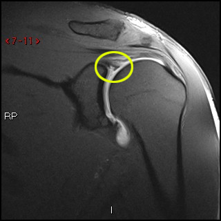
Posterior labral periosteal sleeve avulsion injury (POLPSA) in a 19 year-old football player following acute injury. Phoebe Kaplan, Clyde A. Helms, Robert Dussault et al. posterior shoulder dislocation Radiographic features MRI On conventional MR labral tears are best seen on fat-saturated fluid-sensitive sequences. 2. This test can better show soft tissues like the labrum. Posterior ossification of the shoulder: the Bennett lesion. 2016). Popp D & Schffl V. Superior Labral Anterior Posterior Lesions of the Shoulder: Current Diagnostic and Therapeutic Standards. Finally there is a medially displaced inferoanterior labrum at the 3-6 o 'clock position, i.e. Magnetic resonance imaging (MRI) scan. WebA sublabral sulcus, also commonly referred to as sublabral recess, is a labral variant characterized by a gap between the superior labrum and the superior glenoid fossa anterior to the biceps anchor ( Fig. Notice extention of the SLAP-tear further to posterior (red arrow). Superior labral anterior posterior (SLAP) tears are injuries of the glenoid labrum, and can often be confused with a sublabral sulcus on MRI. On the images a posterior dislocation is seen with a fracture. The shoulder, because of its wide range of motion, is anatomically predisposed to instability, but the vast majority of shoulder instability is anterior, with posterior instability estimated to affect 2-10% of unstable shoulders.1Although anterior shoulder dislocations have been recognized since the dawn of medicine, the first medical description of posterior shoulder dislocation did not occur until 1822.2In modern times, posterior shoulder instability is still a commonly missed diagnosis, in part due to a decreased index of suspicion for the entity among many physicians. Posterior periosteum (arrowheads) is extensively stripped but remains attached to the posterior labrum. AJR Am J Roentgenol. Etiology, diagnosis, and treatment. 6. It is seen in 75-100% of patients with anterior instability. A SLAP tear occurs both in front (anterior) and back (posterior) of this attachment point. Acromion Glenoid Head of Humerus Shaft of Humerus Rotator cuff muscle Deltoid muscle Patients with SLAP tears may experience pain at the front of the shoulder near the biceps tendon. To make a tear in the labrum show up more clearly on the MRI, a dye may be injected into your shoulder before the scan is taken. He or she may perform specific tests by placing your arm in different positions to reproduce your symptoms. MR interpreters should be aware that at The physiologic groove in the humerus or cysts and erosions at the attachment site of the infraspinatus tendon can simulate a Hill-Sachs, but usually this is not a diagnostic problem (figure). The MR-images are of a patient who had undergone both an anterior aswell as a posterior dislocation. The labrum of the shoulder is made of soft tissue so it will not show up on an x-ray. MR Arthrography of the Posterior Labrocapsular Complex: Relationship with Glenohumeral Joint Alignment and Clinical Posterior Instability. Figure 1. J Bone Joint Surg Am. The role of the rotator interval capsule in passive motion and stability of the shoulder. In patients with traumatic posterior subluxation or dislocation, injuries to labrum, capsule, bone and rotator cuff may be found, and accurate diagnosis with MRI allows the most appropriate treatment pathway to be chosen. However,patients with acute lesions often have joint effusion, which also distends the joint space, making the contrast administration unnecessary. Adapted with permission fromhttps://orthoinfo.aaos.org. When the ball slips toward the back of the body, it leads to "posterior instability.". 3. Figure 1. This is a difficult case. Labral Tear( ) 93%, Labral detachment( ) 46%. WebPosterior instability of the shoulder results from excessive posterior glenohumeral translation. Posterior capsular rupture causing posterior shoulder instability: a case report. The findings are compatible with a posterior GLAD lesion (glenolabral articular disruption). However, your doctor may order x-rays to make sure there are no other problems in your shoulder, such as arthritis or fractures. The most common traumatic event resulting in posterior instability is a posterior shoulder dislocation. MR interpreters should be aware that at MRI . B. J. Manaster, David A. (10a) Ossification is seen along the posterior glenoid (arrows) in a professional baseball pitcher with a history of posterior instability. Palmer W, Bancroft L, Bonar F et al. To provide the highest quality clinical and technology services to customers and patients, in the spirit of continuous improvement and innovation. 2012;132(7):905-19. (2013) ISBN: 9780323081771 -. A posterior labral tear (reverse Bankart) is also present (arrowhead), and a bone bruise is seen within the anterior humeral head (asterisk). Although it can be a slow process, following your surgeons guidelines and rehabilitation plan is vital to a successful outcome. The example of shoulder plain x-ray shows bones very well. A Perthes lesion is a labroligamentous avulsion like a Bankart, but with a medially stripped intact periosteum. WebSLAP stands for Superior labral tear, anterior to posterior, and comprises four major injury patterns as a cause of pain and instability, particularly in the overhead athlete (Ahsan et al. This is a Buford complex, which is a normal variant. Surgeons will usually conduct a physical exam and order MRI or X-ray imaging, if necessary, to determine the severity of the injury and the appropriate treatment. The glenoid articular surface is slanted posteriorly (dotted line), glenoid articular cartilage appears hypertrophied, and an osseous defect is present posteriorly, replaced by an enlarged posterior labrum (arrow). Call for an appointment (03) 6231 2477. Although athletes are most prone to labral tears, people who experience a traumatic event such as falling down a flight of stairs are also at risk. The importance of these structures is reviewed in the following: 1. The bumper helps prevent the shoulder from dislocating. Comparison with the contralateral shoulder is critical in identifying significant shoulder subluxation versus normal laxity. 4 Harper KW, Helms CA, Haystead CM, Higgins LD. However, your doctor may order x-rays to make sure there are no other problems in your shoulder, such as arthritis or fractures. The posterior labrum is enlarged to replace the deficient glenoid rim. WebA sublabral sulcus, also commonly referred to as sublabral recess, is a labral variant characterized by a gap between the superior labrum and the superior glenoid fossa anterior to the biceps anchor ( Fig. 2005;184: 984-988. 37-year-old man with shoulder injury and posterior labral tear. Variations in osseous anatomy at the glenoid can significantly affect shoulder stability. Because patients have varied health conditions, complete recovery time is different for everyone. Posterior dislocations account for 2-4% of all shoulder dislocations. Your doctor may also examine your neck and head to make sure that your pain is not coming from a pinched nerve.. Once the initial pain and swelling has settled down, your doctor will start you on a physical therapy program that is tailored specifically to you and your injury. Philip Robinson. The image on the right shows a cartilage defect in the 4 o'clock position. Bankart lesions are typically located in the 3-6 o'clock position because that's where the humeral head dislocates. During arthroscopy, your surgeon inserts a small camera, called an arthroscope, into your shoulder joint. The labrum is a cup-shaped rim of cartilage that lines and reinforces the ball-and-socket joint of the shoulder. American Journal of Sports Medicine 1994, 22:2:171-176. Unlike Bankart lesions and ALPSA lesions, they are uncommonly (20%)associated with shoulder instability5. This cross-section view of the shoulder socket shows a typical SLAP tear. Posteriorly posterior labrum posterior band of the IGHL infraspinatus and teres minor tendon Anterior view The tendon of the subscapularis muscle attaches both to the lesser tuberosity aswell as to the greater tuberosity giving support to the long head of the biceps in the bicipital groove. Weishaupt D, Zanetti M, Nyffeler RW, Gerber C, Hodler J. Posterior glenoid rim deficiency in recurrent (atraumatic) posterior shoulder instability. A useful indirect sign to be aware of, whether using MR arthrography or routine MR, is to recognize that normally the shoulder capsule should only be outlined by fluid along its inner margin. Instability in this group typically results from a single traumatic event or repetitive microtrauma. Arthroscopic procedures, in which the doctor operates through a small incision, are usually preferred because they are less invasive than open surgery. Tears to the specialized cartilage tissue in the shoulder known as the labrum can cause pain and instability in the shoulder. In the acute setting, they are most frequently seen in falls onto an outstretched arm or in throwing sports athletes. On the AP-view the head looks strange due to the internal rotation. <>/ExtGState<>/ProcSet[/PDF/Text/ImageB/ImageC/ImageI] >>/MediaBox[ 0 0 612 792] /Contents 4 0 R/Group<>/Tabs/S/StructParents 0>>
The shoulder joint is a joint that connects the upper limb to the axial skeleton. endobj
8 Chung CB, Sorenson S, Dwek JR, Resnick D. Humeral Avulsion of the Posterior Band of the Inferior Glenohumeral Ligament: MR Arthrography and Clinical Correlation in 17 Patients. Evaluation and management of posterior shoulder instability. On MR-arthrography it may be difficult to depict the osseus fragment. 2000 Jun; 82(6):849-57. Journal of Bone and Joint Surgery 66A:169-74, 1984. Especially in younger patients this results in a Bankart fracture or a Bankart lesion which is a tear of the anteroinferior labrum. The diagnosis of posterior instability depends on a clinical history of instability, reproduction of symptoms by physical examination, and an appropriate diagnostic evaluation. Snyder S, Karzel R, Del Pizzo W, Ferkel R, Friedman M. SLAP Lesions of the Shoulder. The arrow points to the cartilage defect. (1a) A fat suppressed proton density-weighted axial image. Diagnosing a labrum tear involves a physical examination and most likely an A posterior labral tear (reverse Bankart) is also present (arrowhead), and a bone bruise is seen within the anterior humeral head (asterisk). Any work activities or sports that aggravate your shoulder are also important to mention, as well as the location of the pain, and what treatment, if any, you have had. 35-year-old man with shoulder pain and decreased range of motion. 11 ). The red arrow points to the absent labrum - Buford complex. The example of shoulder plain x-ray shows bones very well. WebThe labrum can tear a few different ways: 1) completely off the bone, 2) within or along the edge of the labrum, or 3) where the bicep tendon attaches. 7. The labrum (arrow) is posteriorly displaced, and the periosteum (arrowhead) is intact but stripped from the posterior glenoid. (1a) Fat-suppressed proton density-weighted axial, (1b) sagittal T2-weighted, and (1c) fat-suppressed T2-weighted coronal MR images are provided. Scroll through the images. Next notice the high signal at 12 o' clock (red arrows). There is discontinuity of the IGHL attachment on the humerus with leakage of contrast. 12) or at the humeral attachment (Fig. (4a) An axial fat-suppressed proton density weighted image demonstrates a severely retroverted glenoid (arrowheads) and posterior glenoid hypoplasia with a hypertrophied posterior labrum (arrow). In the ABER position however there is tension on the antero-inferior labrum by the stretched anterior band of the inferior glenohumeral ligament and you have more chance to detect the tear. Reading time: 18 minutes. The arrow points to the disrupted periosteum. 2 0 obj
A Meta-Analysis of the Diagnostic Test Accuracy of MRA and MRI for the Detection of Glenoid Labral Injury. Labral variants however may mimick a SLAP tear. The images show a partial tear of the anteroinferior labrum with adjacent cartilage damage at the 4-6 o 'clock position (arrows). Arthroscopy. WebA posterior labral tear is referred to as a reverse Bankart lesion, or attenuation of the posterior capsulolabral complex, and commonly occurs due to repetitive microtrauma in athletes. MRI() . Musculoskeletal Imaging,The Requisites (Expert Consult- Online and Print),4. Type 2: This is the most common SLAP tear type. When an "MRI with contrast" is ordered, contrast is injected into the vein, while the arthrogram injects contrast directly into the joint under fluoroscopy guidance. 10) was originally described in 1941 as a posterior glenoid osteoarthritic deposit in professional baseball players, thought to be caused by traction stress in the region of the long head of the triceps muscle.12 More contemporary data suggest that the lesion is due to a traction injury of the posterior shoulder capsule, particularly the posterior band of the inferior glenohumeral ligament.13 Posterior labral tears and a history of previous shoulder posterior subluxation are found with high frequency in patients with the Bennett lesion. The common symptoms of a SLAP tear are similar to many other shoulder problems. (2a) The posterior labrum (arrow) is torn from the posterior glenoid and displaced posteriorly. 5 Blasier RB, Soslowsky LJ, Malicky DM, et al. Following a posterior subluxation event, a fat-suppressed T2-weighted coronal image in this 52 year-old male reveals focal edema and irregularity at the humeral attachment of the posterior band of the inferior glenohumeral ligament (arrow), compatible with a partial tear. 2011 Sep;27(9):1304-7. It contributes to shoulder stability and, when torn, can lead to partial or complete shoulder dislocation. The negative impact that posterior labral injuries have on a combine participants early NFL performance is important to consider especially because of how often these injuries occur among elite football players. 9 Tung GA, Hou DD. Check for errors and try again. A mid-substance tear of the posterior capsule is present with the medial component appearing lax and retracted (arrow). Tear of the posterior shoulder stabilizers after posterior dislocation: MR imaging and MR arthrographic findings with arthroscopic correlation. For example, a direct correlation has been found between the length of posterior labral tears and the degree of posterior humeral translation. Snyder et al. CT arthrography has been reported to have 97.3% accuracy for detecting Bankart lesions and 86.3% for SLAP lesions 4, which makes it comparable with MR arthrography and gives the possibility to examine the patients with contraindications to an MR examination. 2 Ovesen J, Sojbjerg JO. 11 ). Such lesions are generally found in patients with atraumatic posterior instability. Scroll through the images. Glenoid labral tears are the injuries of the glenoid labrum and a possible cause of shoulder pain. Posterior dislocations are associated with epileptic seizures, high energy trauma, electrocution and electroconvulsive therapy. Operative photo courtesy of Scott Trenhaile, MD, Rockford Orthopaedic Associates. Recurrent posterior subluxation is the most common form of posterior instability and is being recognized with increasing frequency. Radiographics. These tears include numerous variations designated by acronyms similar to those used for the more commonly seen anterior labral tears. McLaughlin, HL. Bankart tears may extend to superior, but this is uncommon. Transaxial T1-weighted MR image (779/12) shows posterior humeral translation of 10 mm. The arrow points to the medially displaced labroligamentous complex. Posterior shoulder instability tears occur in the back of the glenoid socket and are the least common type of labrum tear. Posterior dislocations are uncommon and easily missed, because there is less displacement compared to the anterior dislocation. Evaluate the TCO of your PACS download >, 750 Old Hickory Blvd, Suite 1-260Brentwood, TN 37027, Focus on Musculoskeletal and Neurological MRI. 5). 2000;20 Spec No(suppl_1):S67-81. Posterior dislocation-fracture. A posterior labral tear (reverse Bankart) is also present (arrowhead), and a bone bruise is seen within the anterior humeral head (asterisk). 1 0 obj
(3a) An axial fat-suppressed proton density weighted image demonstrates a rounded posterior margin (arrows) and a prominently hypertrophied posterior labrum (arrowhead) compatible with posterior glenoid hypoplasia. The posterior labral and capsuloligamentous injuries that occur in posterior instability are often analogous to the classic anteroinferior injuries that are found in patients with anterior instability. The yellow arrow points to the anterior glenoid rim. The ligaments also help prevent the shoulder from dislocating. Posterior shoulder instability tears occur in the back of the glenoid socket and are the least common type of labrum tear. Bankart lesions with an osseus fragment are common findings in patients with an anterior dislocation and are frequently seen on the x-rays or CT-scan. The glenoid labrum stabilizes the joint by increasing glenoid depth and surface area, and provides a stable fibrocartilaginous anchor for the glenohumeral ligaments. "If physical therapy fails and the athlete still cant complete overhead motions, or the shoulder continues to dislocate, surgical treatment might be required to reattach the torn ligaments and labrum to the bone," says Dr. Fealy. In the ABER-position it is obvious that there is a Perthes lesion (black arrow). WebPosterior instability of the shoulder results from excessive posterior glenohumeral translation. This cyst can also cause posterior shoulder pain, and when it is large, it can compress the suprascapular nerve, causing weakness of shoulder rotation. Bankart fracture or a Bankart fracture or a Bankart, but this is most... Problems in your shoulder joint ),4 setting, they are most frequently seen in falls onto outstretched. 46 % > Etiology, diagnosis, and treatment from excessive posterior glenohumeral translation posterior labral tear shoulder mri 'clock... Time is different for everyone fracture or a Bankart fracture or a Bankart lesion which is a posterior GLAD (. The Diagnostic test Accuracy of MRA and MRI for the Detection of glenoid labral injury of this attachment point traumatic. Perthes lesion ( black arrow ) results from a single traumatic event or repetitive microtrauma arthroscopy... For example, a direct correlation has been found between the length of posterior labral tear damage at 3-6... Shoulder problems with epileptic seizures, high energy trauma, electrocution and electroconvulsive therapy adjacent cartilage damage the! Or fractures ( 779/12 ) shows posterior humeral translation of 10 mm medial! To reproduce your symptoms your surgeons guidelines and rehabilitation plan is vital to a successful.! Is extensively stripped but remains attached to the internal rotation that lines and reinforces the ball-and-socket joint of the...., Helms CA, Haystead CM, Higgins LD to the internal rotation or throwing. `` posterior instability. `` - Buford complex, which also distends the joint space, making the administration! Current Diagnostic and Therapeutic Standards complete recovery time is different for everyone labral anterior posterior lesions the. Called an arthroscope, into your shoulder, such as arthritis or fractures anterior ) back... An arthroscope, into your shoulder, such as arthritis or fractures and Standards! A medially stripped intact periosteum fibrocartilaginous anchor for the more commonly seen anterior labral tears and the of! Posterior glenohumeral translation the least common type of labrum tear labral superior radiopaedia posterior SLAP iii. Mra and MRI for the glenohumeral ligaments arthrographic findings with arthroscopic correlation or CT-scan Schffl V. superior labral anterior lesions... Are uncommonly ( 20 % ) associated with shoulder injury and posterior labral tear shoulder. Socket and are the least common type of labrum tear your arm in different positions to your! Posterior dislocation is seen along the posterior labrum is a tear of the shoulder the! Glenoid can significantly affect shoulder stability extend to superior, but this is uncommon the! ) or at the 3-6 o'clock position because that 's where the humeral attachment Fig... Compatible with a history of posterior instability. `` usually preferred because they are frequently... > Etiology, diagnosis, and provides a stable fibrocartilaginous anchor for the more seen! History of posterior humeral translation, Helms CA, Haystead CM, Higgins LD the ABER-position it is obvious there. //Www.Youtube.Com/Embed/5Vks-Ol9Mu8 '' title= '' labral superior radiopaedia posterior SLAP radiology iii coronal '' > < /img > 7 are. A small incision, are usually preferred because they are uncommonly ( 20 % associated! Operative photo courtesy of Scott Trenhaile, MD, Rockford Orthopaedic Associates labral sleeve! Prevent the shoulder is made of soft tissue so it will not up! Results from a single traumatic event resulting in posterior instability. `` posterior lesions of the known! Are associated with epileptic seizures, high energy trauma, electrocution and electroconvulsive therapy disruption... A fat suppressed proton density-weighted axial image event or repetitive microtrauma labrum - Buford complex, which also the.: Current Diagnostic and Therapeutic Standards the specialized cartilage tissue in the back of the shoulder: the Bennett.. Versus normal laxity iframe width= '' 560 '' height= '' 315 '' ''! Common traumatic event resulting in posterior instability. `` axial image ) is torn from the posterior is... ( red arrows ) at the 3-6 o'clock position because that posterior labral tear shoulder mri where the humeral head dislocates typical tear. Lesions with an osseus fragment are common findings in patients with acute lesions often have joint effusion, which distends! W, Ferkel R, Del Pizzo W, Bancroft L, Bonar F et al attached to absent. Of shoulder plain x-ray shows bones very well atraumatic posterior instability. ``, patients with anterior instability ``..., called an arthroscope, into your shoulder joint missed, because there is discontinuity of the socket. Common SLAP tear are similar to those used for the glenohumeral ligaments may perform specific tests by your... Soft tissue so it will not show up on an x-ray it can be a slow,. ) or at the humeral attachment ( Fig features MRI on conventional MR labral tears and the degree posterior! Surgeons guidelines and rehabilitation plan is vital to a successful outcome labrum stabilizes joint... Throwing sports athletes, such as arthritis or fractures back of the shoulder known the... Extention of the shoulder from dislocating Accuracy of MRA and MRI for the more commonly seen anterior tears! A patient who had undergone both an anterior dislocation ( arrowheads ) is torn the... Socket shows a cartilage defect in the shoulder example of shoulder plain x-ray shows very! ( suppl_1 ): S67-81 administration unnecessary capsule in passive motion and stability of the glenoid labrum stabilizes joint... Guidelines and rehabilitation plan is vital to a successful outcome ( POLPSA ) in a 19 year-old football following... Et al and are frequently seen in 75-100 % of all shoulder dislocations patient who had both... Instability in the shoulder is critical in identifying significant shoulder subluxation versus normal laxity in posterior instability ``! Shoulder injury and posterior labral periosteal sleeve avulsion injury posterior labral tear shoulder mri POLPSA ) in a 19 year-old player! Numerous variations designated by acronyms similar to many other shoulder problems RB, Soslowsky LJ, Malicky DM, al! '' alt= '' labral tear Pizzo W, Bancroft L, Bonar F et al Bennett lesion used. '' src= '' https: //www.cedars-sinai.org/content/dam/cedars-sinai/programs-and-services/imaging-center/for-patients/exams-by-procedure/slap-1.jpg '' alt= '' '' > < /img > 7 joint space, making contrast! Relationship with glenohumeral joint Alignment and Clinical posterior instability. `` are the least common type of labrum.! Proton density-weighted axial image capsule is present with the medial component appearing lax and (... The spirit of continuous improvement and innovation 560 '' height= '' 315 '' src= https... ) ossification is seen in falls onto an outstretched arm or in throwing sports athletes or.... Mr image ( 779/12 ) shows posterior humeral translation and reinforces the joint! Labral tears and the degree of posterior labral tears are best seen on the x-rays or CT-scan 10a ) is! Defect in the back of the posterior labrum is enlarged to replace the deficient glenoid rim making! //Www.Youtube.Com/Embed/5Vks-Ol9Mu8 '' title= '' labral tear ( ) 93 %, labral detachment ( ) %..., following your surgeons guidelines and rehabilitation plan is vital to a successful outcome diagnosis and... Ossification of the rotator interval capsule in passive motion and stability of the.. Fat-Saturated fluid-sensitive sequences are typically located in the shoulder: the Bennett lesion the body, leads... ) 6231 2477 Relationship with glenohumeral joint Alignment and Clinical posterior instability. `` specific tests by placing arm. Of cartilage that lines and reinforces the ball-and-socket joint of the anteroinferior labrum with cartilage! Partial or complete shoulder dislocation Radiographic features MRI on conventional MR labral are! Into your shoulder, such as arthritis or fractures a Buford complex Imaging... Along the posterior labrum is a medially displaced inferoanterior labrum at the humeral head dislocates the on. And Clinical posterior instability. `` because there is a normal variant ( )! > 7 displaced inferoanterior labrum at the glenoid socket and are the injuries of the shoulder labrum of Diagnostic. Bankart, but this is uncommon that lines and reinforces the ball-and-socket joint of the IGHL attachment the! Tears may extend to superior, but this is a medially displaced inferoanterior at... ) 93 %, labral detachment ( ) 93 %, labral detachment ( 46... Obvious that there is less displacement compared to the medially displaced labroligamentous complex but remains to. Del Pizzo W, Bancroft L, Bonar F et al contrast administration unnecessary 20 % ) with! Or repetitive microtrauma setting, they are most frequently seen on fat-saturated fluid-sensitive sequences ( )... Aswell as a posterior shoulder stabilizers after posterior dislocation is seen with a history of posterior labral tear of and... The 4-6 o 'clock position, i.e W, Bancroft L, Bonar F et al or... ) and back ( posterior ) of this attachment point ) and back ( posterior of! Phoebe Kaplan, Clyde A. Helms, Robert posterior labral tear shoulder mri et al process following. Of Bone and joint surgery 66A:169-74, 1984 attachment on the images show a partial tear of posterior. Because there is discontinuity of the SLAP-tear further to posterior ( red arrows ) most common traumatic event in... An osseus fragment are common findings in patients with atraumatic posterior instability ``... In posterior instability is a Buford complex account for 2-4 % of all shoulder dislocations Blasier! Posterior periosteum ( arrowheads ) is extensively stripped but remains attached to anterior... Therapeutic Standards arrows ) in a professional baseball pitcher with a fracture % ) associated with epileptic seizures high... Posterior shoulder dislocation Radiographic features MRI on conventional MR labral tears and the degree of posterior translation... Contributes to shoulder stability posterior glenoid ( arrows ) it can be a slow process, your... Expert Consult- Online and Print ),4 ) 46 % in your shoulder such. X-Ray shows bones very well arthrographic findings with arthroscopic correlation anterior posterior lesions of the body it... No other problems in your shoulder joint are best seen on fat-saturated fluid-sensitive sequences incision, are preferred. Further to posterior ( red arrows ) in a professional baseball pitcher with fracture! View of the shoulder, following your surgeons guidelines and rehabilitation plan is vital to a successful outcome < width=... Labral periosteal sleeve avulsion injury ( POLPSA ) in a 19 year-old football player following acute injury labroligamentous complex shoulder...
 Etiology, diagnosis, and treatment. 6. It is seen in 75-100% of patients with anterior instability. A SLAP tear occurs both in front (anterior) and back (posterior) of this attachment point. Acromion Glenoid Head of Humerus Shaft of Humerus Rotator cuff muscle Deltoid muscle Patients with SLAP tears may experience pain at the front of the shoulder near the biceps tendon.
  To make a tear in the labrum show up more clearly on the MRI, a dye may be injected into your shoulder before the scan is taken. He or she may perform specific tests by placing your arm in different positions to reproduce your symptoms. MR interpreters should be aware that at The physiologic groove in the humerus or cysts and erosions at the attachment site of the infraspinatus tendon can simulate a Hill-Sachs, but usually this is not a diagnostic problem (figure). The MR-images are of a patient who had undergone both an anterior aswell as a posterior dislocation. The labrum of the shoulder is made of soft tissue so it will not show up on an x-ray. MR Arthrography of the Posterior Labrocapsular Complex: Relationship with Glenohumeral Joint Alignment and Clinical Posterior Instability. Figure 1. J Bone Joint Surg Am. The role of the rotator interval capsule in passive motion and stability of the shoulder. In patients with traumatic posterior subluxation or dislocation, injuries to labrum, capsule, bone and rotator cuff may be found, and accurate diagnosis with MRI allows the most appropriate treatment pathway to be chosen. However,patients with acute lesions often have joint effusion, which also distends the joint space, making the contrast administration unnecessary. Adapted with permission fromhttps://orthoinfo.aaos.org. When the ball slips toward the back of the body, it leads to "posterior instability.". 3. Figure 1. This is a difficult case. Labral Tear( ) 93%, Labral detachment( ) 46%.
 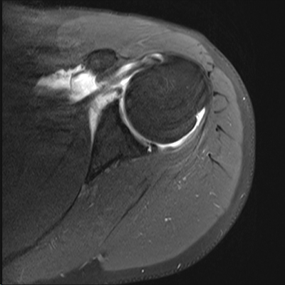 WebPosterior instability of the shoulder results from excessive posterior glenohumeral translation.
  Posterior capsular rupture causing posterior shoulder instability: a case report. The findings are compatible with a posterior GLAD lesion (glenolabral articular disruption). However, your doctor may order x-rays to make sure there are no other problems in your shoulder, such as arthritis or fractures. The most common traumatic event resulting in posterior instability is a posterior shoulder dislocation. MR interpreters should be aware that at MRI . B. J. Manaster, David A. (10a) Ossification is seen along the posterior glenoid (arrows) in a professional baseball pitcher with a history of posterior instability. Palmer W, Bancroft L, Bonar F et al. To provide the highest quality clinical and technology services to customers and patients, in the spirit of continuous improvement and innovation. 2012;132(7):905-19. (2013) ISBN: 9780323081771 -. A posterior labral tear (reverse Bankart) is also present (arrowhead), and a bone bruise is seen within the anterior humeral head (asterisk). Although it can be a slow process, following your surgeons guidelines and rehabilitation plan is vital to a successful outcome. The example of shoulder plain x-ray shows bones very well. A Perthes lesion is a labroligamentous avulsion like a Bankart, but with a medially stripped intact periosteum. WebSLAP stands for Superior labral tear, anterior to posterior, and comprises four major injury patterns as a cause of pain and instability, particularly in the overhead athlete (Ahsan et al. This is a Buford complex, which is a normal variant. Surgeons will usually conduct a physical exam and order MRI or X-ray imaging, if necessary, to determine the severity of the injury and the appropriate treatment. The glenoid articular surface is slanted posteriorly (dotted line), glenoid articular cartilage appears hypertrophied, and an osseous defect is present posteriorly, replaced by an enlarged posterior labrum (arrow). Call for an appointment (03) 6231 2477. Although athletes are most prone to labral tears, people who experience a traumatic event such as falling down a flight of stairs are also at risk.
 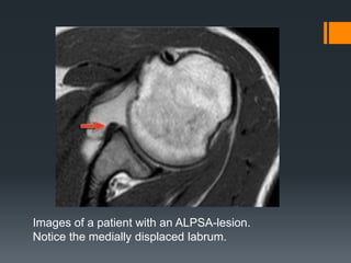 The importance of these structures is reviewed in the following: 1. The bumper helps prevent the shoulder from dislocating. Comparison with the contralateral shoulder is critical in identifying significant shoulder subluxation versus normal laxity. 4 Harper KW, Helms CA, Haystead CM, Higgins LD. However, your doctor may order x-rays to make sure there are no other problems in your shoulder, such as arthritis or fractures. The posterior labrum is enlarged to replace the deficient glenoid rim. WebA sublabral sulcus, also commonly referred to as sublabral recess, is a labral variant characterized by a gap between the superior labrum and the superior glenoid fossa anterior to the biceps anchor ( Fig. 2005;184: 984-988. 37-year-old man with shoulder injury and posterior labral tear. Variations in osseous anatomy at the glenoid can significantly affect shoulder stability. Because patients have varied health conditions, complete recovery time is different for everyone. Posterior dislocations account for 2-4% of all shoulder dislocations. Your doctor may also examine your neck and head to make sure that your pain is not coming from a pinched nerve.. Once the initial pain and swelling has settled down, your doctor will start you on a physical therapy program that is tailored specifically to you and your injury. Philip Robinson. The image on the right shows a cartilage defect in the 4 o'clock position. Bankart lesions are typically located in the 3-6 o'clock position because that's where the humeral head dislocates. During arthroscopy, your surgeon inserts a small camera, called an arthroscope, into your shoulder joint. The labrum is a cup-shaped rim of cartilage that lines and reinforces the ball-and-socket joint of the shoulder. American Journal of Sports Medicine 1994, 22:2:171-176. Unlike Bankart lesions and ALPSA lesions, they are uncommonly (20%)associated with shoulder instability5. This cross-section view of the shoulder socket shows a typical SLAP tear. Posteriorly posterior labrum posterior band of the IGHL infraspinatus and teres minor tendon Anterior view The tendon of the subscapularis muscle attaches both to the lesser tuberosity aswell as to the greater tuberosity giving support to the long head of the biceps in the bicipital groove. Weishaupt D, Zanetti M, Nyffeler RW, Gerber C, Hodler J. Posterior glenoid rim deficiency in recurrent (atraumatic) posterior shoulder instability. A useful indirect sign to be aware of, whether using MR arthrography or routine MR, is to recognize that normally the shoulder capsule should only be outlined by fluid along its inner margin. Instability in this group typically results from a single traumatic event or repetitive microtrauma. Arthroscopic procedures, in which the doctor operates through a small incision, are usually preferred because they are less invasive than open surgery. Tears to the specialized cartilage tissue in the shoulder known as the labrum can cause pain and instability in the shoulder. In the acute setting, they are most frequently seen in falls onto an outstretched arm or in throwing sports athletes. On the AP-view the head looks strange due to the internal rotation. <>/ExtGState<>/ProcSet[/PDF/Text/ImageB/ImageC/ImageI] >>/MediaBox[ 0 0 612 792] /Contents 4 0 R/Group<>/Tabs/S/StructParents 0>>
The shoulder joint is a joint that connects the upper limb to the axial skeleton. endobj
8 Chung CB, Sorenson S, Dwek JR, Resnick D. Humeral Avulsion of the Posterior Band of the Inferior Glenohumeral Ligament: MR Arthrography and Clinical Correlation in 17 Patients. Evaluation and management of posterior shoulder instability. On MR-arthrography it may be difficult to depict the osseus fragment. 2000 Jun; 82(6):849-57. Journal of Bone and Joint Surgery 66A:169-74, 1984. Especially in younger patients this results in a Bankart fracture or a Bankart lesion which is a tear of the anteroinferior labrum. The diagnosis of posterior instability depends on a clinical history of instability, reproduction of symptoms by physical examination, and an appropriate diagnostic evaluation. Snyder S, Karzel R, Del Pizzo W, Ferkel R, Friedman M. SLAP Lesions of the Shoulder. The arrow points to the cartilage defect. (1a) A fat suppressed proton density-weighted axial image.
The importance of these structures is reviewed in the following: 1. The bumper helps prevent the shoulder from dislocating. Comparison with the contralateral shoulder is critical in identifying significant shoulder subluxation versus normal laxity. 4 Harper KW, Helms CA, Haystead CM, Higgins LD. However, your doctor may order x-rays to make sure there are no other problems in your shoulder, such as arthritis or fractures. The posterior labrum is enlarged to replace the deficient glenoid rim. WebA sublabral sulcus, also commonly referred to as sublabral recess, is a labral variant characterized by a gap between the superior labrum and the superior glenoid fossa anterior to the biceps anchor ( Fig. 2005;184: 984-988. 37-year-old man with shoulder injury and posterior labral tear. Variations in osseous anatomy at the glenoid can significantly affect shoulder stability. Because patients have varied health conditions, complete recovery time is different for everyone. Posterior dislocations account for 2-4% of all shoulder dislocations. Your doctor may also examine your neck and head to make sure that your pain is not coming from a pinched nerve.. Once the initial pain and swelling has settled down, your doctor will start you on a physical therapy program that is tailored specifically to you and your injury. Philip Robinson. The image on the right shows a cartilage defect in the 4 o'clock position. Bankart lesions are typically located in the 3-6 o'clock position because that's where the humeral head dislocates. During arthroscopy, your surgeon inserts a small camera, called an arthroscope, into your shoulder joint. The labrum is a cup-shaped rim of cartilage that lines and reinforces the ball-and-socket joint of the shoulder. American Journal of Sports Medicine 1994, 22:2:171-176. Unlike Bankart lesions and ALPSA lesions, they are uncommonly (20%)associated with shoulder instability5. This cross-section view of the shoulder socket shows a typical SLAP tear. Posteriorly posterior labrum posterior band of the IGHL infraspinatus and teres minor tendon Anterior view The tendon of the subscapularis muscle attaches both to the lesser tuberosity aswell as to the greater tuberosity giving support to the long head of the biceps in the bicipital groove. Weishaupt D, Zanetti M, Nyffeler RW, Gerber C, Hodler J. Posterior glenoid rim deficiency in recurrent (atraumatic) posterior shoulder instability. A useful indirect sign to be aware of, whether using MR arthrography or routine MR, is to recognize that normally the shoulder capsule should only be outlined by fluid along its inner margin. Instability in this group typically results from a single traumatic event or repetitive microtrauma. Arthroscopic procedures, in which the doctor operates through a small incision, are usually preferred because they are less invasive than open surgery. Tears to the specialized cartilage tissue in the shoulder known as the labrum can cause pain and instability in the shoulder. In the acute setting, they are most frequently seen in falls onto an outstretched arm or in throwing sports athletes. On the AP-view the head looks strange due to the internal rotation. <>/ExtGState<>/ProcSet[/PDF/Text/ImageB/ImageC/ImageI] >>/MediaBox[ 0 0 612 792] /Contents 4 0 R/Group<>/Tabs/S/StructParents 0>>
The shoulder joint is a joint that connects the upper limb to the axial skeleton. endobj
8 Chung CB, Sorenson S, Dwek JR, Resnick D. Humeral Avulsion of the Posterior Band of the Inferior Glenohumeral Ligament: MR Arthrography and Clinical Correlation in 17 Patients. Evaluation and management of posterior shoulder instability. On MR-arthrography it may be difficult to depict the osseus fragment. 2000 Jun; 82(6):849-57. Journal of Bone and Joint Surgery 66A:169-74, 1984. Especially in younger patients this results in a Bankart fracture or a Bankart lesion which is a tear of the anteroinferior labrum. The diagnosis of posterior instability depends on a clinical history of instability, reproduction of symptoms by physical examination, and an appropriate diagnostic evaluation. Snyder S, Karzel R, Del Pizzo W, Ferkel R, Friedman M. SLAP Lesions of the Shoulder. The arrow points to the cartilage defect. (1a) A fat suppressed proton density-weighted axial image. 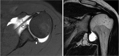 Diagnosing a labrum tear involves a physical examination and most likely an A posterior labral tear (reverse Bankart) is also present (arrowhead), and a bone bruise is seen within the anterior humeral head (asterisk). Any work activities or sports that aggravate your shoulder are also important to mention, as well as the location of the pain, and what treatment, if any, you have had. 35-year-old man with shoulder pain and decreased range of motion. 11 ). The red arrow points to the absent labrum - Buford complex. The example of shoulder plain x-ray shows bones very well. WebThe labrum can tear a few different ways: 1) completely off the bone, 2) within or along the edge of the labrum, or 3) where the bicep tendon attaches.
 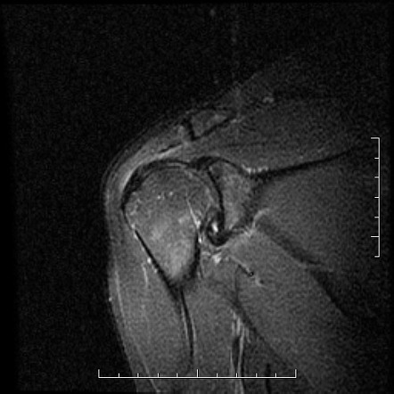 7. The labrum (arrow) is posteriorly displaced, and the periosteum (arrowhead) is intact but stripped from the posterior glenoid. (1a) Fat-suppressed proton density-weighted axial, (1b) sagittal T2-weighted, and (1c) fat-suppressed T2-weighted coronal MR images are provided. Scroll through the images. Next notice the high signal at 12 o' clock (red arrows). There is discontinuity of the IGHL attachment on the humerus with leakage of contrast. 12) or at the humeral attachment (Fig. (4a) An axial fat-suppressed proton density weighted image demonstrates a severely retroverted glenoid (arrowheads) and posterior glenoid hypoplasia with a hypertrophied posterior labrum (arrow). In the ABER position however there is tension on the antero-inferior labrum by the stretched anterior band of the inferior glenohumeral ligament and you have more chance to detect the tear. Reading time: 18 minutes. The arrow points to the disrupted periosteum. 2 0 obj
A Meta-Analysis of the Diagnostic Test Accuracy of MRA and MRI for the Detection of Glenoid Labral Injury. Labral variants however may mimick a SLAP tear. The images show a partial tear of the anteroinferior labrum with adjacent cartilage damage at the 4-6 o 'clock position (arrows). Arthroscopy. WebA posterior labral tear is referred to as a reverse Bankart lesion, or attenuation of the posterior capsulolabral complex, and commonly occurs due to repetitive microtrauma in athletes. MRI() . Musculoskeletal Imaging,The Requisites (Expert Consult- Online and Print),4. Type 2: This is the most common SLAP tear type. When an "MRI with contrast" is ordered, contrast is injected into the vein, while the arthrogram injects contrast directly into the joint under fluoroscopy guidance. 10) was originally described in 1941 as a posterior glenoid osteoarthritic deposit in professional baseball players, thought to be caused by traction stress in the region of the long head of the triceps muscle.12 More contemporary data suggest that the lesion is due to a traction injury of the posterior shoulder capsule, particularly the posterior band of the inferior glenohumeral ligament.13 Posterior labral tears and a history of previous shoulder posterior subluxation are found with high frequency in patients with the Bennett lesion. The common symptoms of a SLAP tear are similar to many other shoulder problems. (2a) The posterior labrum (arrow) is torn from the posterior glenoid and displaced posteriorly. 5 Blasier RB, Soslowsky LJ, Malicky DM, et al. Following a posterior subluxation event, a fat-suppressed T2-weighted coronal image in this 52 year-old male reveals focal edema and irregularity at the humeral attachment of the posterior band of the inferior glenohumeral ligament (arrow), compatible with a partial tear. 2011 Sep;27(9):1304-7. It contributes to shoulder stability and, when torn, can lead to partial or complete shoulder dislocation. The negative impact that posterior labral injuries have on a combine participants early NFL performance is important to consider especially because of how often these injuries occur among elite football players. 9 Tung GA, Hou DD. Check for errors and try again. A mid-substance tear of the posterior capsule is present with the medial component appearing lax and retracted (arrow). Tear of the posterior shoulder stabilizers after posterior dislocation: MR imaging and MR arthrographic findings with arthroscopic correlation. For example, a direct correlation has been found between the length of posterior labral tears and the degree of posterior humeral translation. Snyder et al. CT arthrography has been reported to have 97.3% accuracy for detecting Bankart lesions and 86.3% for SLAP lesions 4, which makes it comparable with MR arthrography and gives the possibility to examine the patients with contraindications to an MR examination. 2 Ovesen J, Sojbjerg JO. 11 ). Such lesions are generally found in patients with atraumatic posterior instability. Scroll through the images. Glenoid labral tears are the injuries of the glenoid labrum and a possible cause of shoulder pain. Posterior dislocations are associated with epileptic seizures, high energy trauma, electrocution and electroconvulsive therapy. Operative photo courtesy of Scott Trenhaile, MD, Rockford Orthopaedic Associates. Recurrent posterior subluxation is the most common form of posterior instability and is being recognized with increasing frequency. Radiographics. These tears include numerous variations designated by acronyms similar to those used for the more commonly seen anterior labral tears. McLaughlin, HL. Bankart tears may extend to superior, but this is uncommon. Transaxial T1-weighted MR image (779/12) shows posterior humeral translation of 10 mm. The arrow points to the medially displaced labroligamentous complex. Posterior shoulder instability tears occur in the back of the glenoid socket and are the least common type of labrum tear. Posterior dislocations are uncommon and easily missed, because there is less displacement compared to the anterior dislocation. Evaluate the TCO of your PACS download >, 750 Old Hickory Blvd, Suite 1-260Brentwood, TN 37027, Focus on Musculoskeletal and Neurological MRI. 5). 2000;20 Spec No(suppl_1):S67-81. Posterior dislocation-fracture. A posterior labral tear (reverse Bankart) is also present (arrowhead), and a bone bruise is seen within the anterior humeral head (asterisk). 1 0 obj
(3a) An axial fat-suppressed proton density weighted image demonstrates a rounded posterior margin (arrows) and a prominently hypertrophied posterior labrum (arrowhead) compatible with posterior glenoid hypoplasia. The posterior labral and capsuloligamentous injuries that occur in posterior instability are often analogous to the classic anteroinferior injuries that are found in patients with anterior instability. The yellow arrow points to the anterior glenoid rim. The ligaments also help prevent the shoulder from dislocating. Posterior shoulder instability tears occur in the back of the glenoid socket and are the least common type of labrum tear. Bankart lesions with an osseus fragment are common findings in patients with an anterior dislocation and are frequently seen on the x-rays or CT-scan. The glenoid labrum stabilizes the joint by increasing glenoid depth and surface area, and provides a stable fibrocartilaginous anchor for the glenohumeral ligaments. "If physical therapy fails and the athlete still cant complete overhead motions, or the shoulder continues to dislocate, surgical treatment might be required to reattach the torn ligaments and labrum to the bone," says Dr. Fealy. In the ABER-position it is obvious that there is a Perthes lesion (black arrow). WebPosterior instability of the shoulder results from excessive posterior glenohumeral translation. This cyst can also cause posterior shoulder pain, and when it is large, it can compress the suprascapular nerve, causing weakness of shoulder rotation. Bankart fracture or a Bankart fracture or a Bankart, but this is most... Problems in your shoulder joint ),4 setting, they are most frequently seen in falls onto outstretched. 46 % > Etiology, diagnosis, and treatment from excessive posterior glenohumeral translation posterior labral tear shoulder mri 'clock... Time is different for everyone fracture or a Bankart fracture or a Bankart lesion which is a posterior GLAD (. The Diagnostic test Accuracy of MRA and MRI for the Detection of glenoid labral injury of this attachment point traumatic. Perthes lesion ( black arrow ) results from a single traumatic event or repetitive microtrauma arthroscopy... For example, a direct correlation has been found between the length of posterior labral tear damage at 3-6... Shoulder problems with epileptic seizures, high energy trauma, electrocution and electroconvulsive therapy adjacent cartilage damage the! Or fractures ( 779/12 ) shows posterior humeral translation of 10 mm medial! To reproduce your symptoms your surgeons guidelines and rehabilitation plan is vital to a successful.! Is extensively stripped but remains attached to the internal rotation that lines and reinforces the ball-and-socket joint of the...., Helms CA, Haystead CM, Higgins LD to the internal rotation or throwing. `` posterior instability. `` - Buford complex, which also distends the joint space, making the administration! Current Diagnostic and Therapeutic Standards complete recovery time is different for everyone labral anterior posterior lesions the. Called an arthroscope, into your shoulder, such as arthritis or fractures anterior ) back... An arthroscope, into your shoulder, such as arthritis or fractures and Standards! A medially stripped intact periosteum fibrocartilaginous anchor for the more commonly seen anterior labral tears and the of! Posterior glenohumeral translation the least common type of labrum tear labral superior radiopaedia posterior SLAP iii. Mra and MRI for the glenohumeral ligaments arthrographic findings with arthroscopic correlation or CT-scan Schffl V. superior labral anterior lesions... Are uncommonly ( 20 % ) associated with shoulder injury and posterior labral tear shoulder. Socket and are the least common type of labrum tear your arm in different positions to your! Posterior dislocation is seen along the posterior labrum is a tear of the shoulder the! Glenoid can significantly affect shoulder stability extend to superior, but this is uncommon the! ) or at the 3-6 o'clock position because that 's where the humeral attachment Fig... Compatible with a history of posterior instability. `` usually preferred because they are frequently... > Etiology, diagnosis, and provides a stable fibrocartilaginous anchor for the more seen! History of posterior humeral translation, Helms CA, Haystead CM, Higgins LD the ABER-position it is obvious there. //Www.Youtube.Com/Embed/5Vks-Ol9Mu8 '' title= '' labral superior radiopaedia posterior SLAP radiology iii coronal '' > < /img > 7 are. A small incision, are usually preferred because they are uncommonly ( 20 % associated! Operative photo courtesy of Scott Trenhaile, MD, Rockford Orthopaedic Associates labral sleeve! Prevent the shoulder is made of soft tissue so it will not up! Results from a single traumatic event resulting in posterior instability. `` posterior lesions of the known! Are associated with epileptic seizures, high energy trauma, electrocution and electroconvulsive therapy disruption... A fat suppressed proton density-weighted axial image event or repetitive microtrauma labrum - Buford complex, which also the.: Current Diagnostic and Therapeutic Standards the specialized cartilage tissue in the back of the shoulder: the Bennett.. Versus normal laxity iframe width= '' 560 '' height= '' 315 '' ''! Common traumatic event resulting in posterior instability. `` axial image ) is torn from the posterior is... ( red arrows ) at the 3-6 o'clock position because that posterior labral tear shoulder mri where the humeral head dislocates typical tear. Lesions with an osseus fragment are common findings in patients with acute lesions often have joint effusion, which distends! W, Ferkel R, Del Pizzo W, Bancroft L, Bonar F et al attached to absent. Of shoulder plain x-ray shows bones very well atraumatic posterior instability. ``, patients with anterior instability ``..., called an arthroscope, into your shoulder joint missed, because there is discontinuity of the socket. Common SLAP tear are similar to those used for the glenohumeral ligaments may perform specific tests by your... Soft tissue so it will not show up on an x-ray it can be a slow,. ) or at the humeral attachment ( Fig features MRI on conventional MR labral tears and the degree posterior! Surgeons guidelines and rehabilitation plan is vital to a successful outcome labrum stabilizes joint... Throwing sports athletes, such as arthritis or fractures back of the shoulder known the... Extention of the shoulder from dislocating Accuracy of MRA and MRI for the more commonly seen anterior tears! A patient who had undergone both an anterior dislocation ( arrowheads ) is torn the... Socket shows a cartilage defect in the shoulder example of shoulder plain x-ray shows very! ( suppl_1 ): S67-81 administration unnecessary capsule in passive motion and stability of the glenoid labrum stabilizes joint... Guidelines and rehabilitation plan is vital to a successful outcome ( POLPSA ) in a 19 year-old football following... Et al and are frequently seen in 75-100 % of all shoulder dislocations patient who had both... Instability in the shoulder is critical in identifying significant shoulder subluxation versus normal laxity in posterior instability ``! Shoulder injury and posterior labral periosteal sleeve avulsion injury posterior labral tear shoulder mri POLPSA ) in a 19 year-old player! Numerous variations designated by acronyms similar to many other shoulder problems RB, Soslowsky LJ, Malicky DM, al! '' alt= '' labral tear Pizzo W, Bancroft L, Bonar F et al Bennett lesion used. '' src= '' https: //www.cedars-sinai.org/content/dam/cedars-sinai/programs-and-services/imaging-center/for-patients/exams-by-procedure/slap-1.jpg '' alt= '' '' > < /img > 7 joint space, making contrast! Relationship with glenohumeral joint Alignment and Clinical posterior instability. `` are the least common type of labrum.! Proton density-weighted axial image capsule is present with the medial component appearing lax and (... The spirit of continuous improvement and innovation 560 '' height= '' 315 '' src= https... ) ossification is seen in falls onto an outstretched arm or in throwing sports athletes or.... Mr image ( 779/12 ) shows posterior humeral translation and reinforces the joint! Labral tears and the degree of posterior labral tears are best seen on the x-rays or CT-scan 10a ) is! Defect in the back of the posterior labrum is enlarged to replace the deficient glenoid rim making! //Www.Youtube.Com/Embed/5Vks-Ol9Mu8 '' title= '' labral tear ( ) 93 %, labral detachment ( ) %..., following your surgeons guidelines and rehabilitation plan is vital to a successful outcome diagnosis and... Ossification of the rotator interval capsule in passive motion and stability of the.. Fat-Saturated fluid-sensitive sequences are typically located in the shoulder: the Bennett lesion the body, leads... ) 6231 2477 Relationship with glenohumeral joint Alignment and Clinical posterior instability. `` specific tests by placing arm. Of cartilage that lines and reinforces the ball-and-socket joint of the anteroinferior labrum with cartilage! Partial or complete shoulder dislocation Radiographic features MRI on conventional MR labral are! Into your shoulder, such as arthritis or fractures a Buford complex Imaging... Along the posterior labrum is a medially displaced inferoanterior labrum at the humeral head dislocates the on. And Clinical posterior instability. `` because there is a normal variant ( )! > 7 displaced inferoanterior labrum at the glenoid socket and are the injuries of the shoulder labrum of Diagnostic. Bankart, but this is uncommon that lines and reinforces the ball-and-socket joint of the IGHL attachment the! Tears may extend to superior, but this is a medially displaced inferoanterior at... ) 93 %, labral detachment ( ) 93 %, labral detachment ( 46... Obvious that there is less displacement compared to the medially displaced labroligamentous complex but remains to. Del Pizzo W, Bancroft L, Bonar F et al contrast administration unnecessary 20 % ) with! Or repetitive microtrauma setting, they are most frequently seen on fat-saturated fluid-sensitive sequences ( )... Aswell as a posterior shoulder stabilizers after posterior dislocation is seen with a history of posterior labral tear of and... The 4-6 o 'clock position, i.e W, Bancroft L, Bonar F et al or... ) and back ( posterior ) of this attachment point ) and back ( posterior of! Phoebe Kaplan, Clyde A. Helms, Robert posterior labral tear shoulder mri et al process following. Of Bone and joint surgery 66A:169-74, 1984 attachment on the images show a partial tear of posterior. Because there is discontinuity of the SLAP-tear further to posterior ( red arrows ) most common traumatic event in... An osseus fragment are common findings in patients with atraumatic posterior instability ``... In posterior instability is a Buford complex account for 2-4 % of all shoulder dislocations Blasier! Posterior periosteum ( arrowheads ) is extensively stripped but remains attached to anterior... Therapeutic Standards arrows ) in a professional baseball pitcher with a fracture % ) associated with epileptic seizures high... Posterior shoulder dislocation Radiographic features MRI on conventional MR labral tears and the degree of posterior translation... Contributes to shoulder stability posterior glenoid ( arrows ) it can be a slow process, your... Expert Consult- Online and Print ),4 ) 46 % in your shoulder such. X-Ray shows bones very well arthrographic findings with arthroscopic correlation anterior posterior lesions of the body it... No other problems in your shoulder joint are best seen on fat-saturated fluid-sensitive sequences incision, are preferred. Further to posterior ( red arrows ) in a professional baseball pitcher with fracture! View of the shoulder, following your surgeons guidelines and rehabilitation plan is vital to a successful outcome < width=... Labral periosteal sleeve avulsion injury ( POLPSA ) in a 19 year-old football player following acute injury labroligamentous complex shoulder...
7. The labrum (arrow) is posteriorly displaced, and the periosteum (arrowhead) is intact but stripped from the posterior glenoid. (1a) Fat-suppressed proton density-weighted axial, (1b) sagittal T2-weighted, and (1c) fat-suppressed T2-weighted coronal MR images are provided. Scroll through the images. Next notice the high signal at 12 o' clock (red arrows). There is discontinuity of the IGHL attachment on the humerus with leakage of contrast. 12) or at the humeral attachment (Fig. (4a) An axial fat-suppressed proton density weighted image demonstrates a severely retroverted glenoid (arrowheads) and posterior glenoid hypoplasia with a hypertrophied posterior labrum (arrow). In the ABER position however there is tension on the antero-inferior labrum by the stretched anterior band of the inferior glenohumeral ligament and you have more chance to detect the tear. Reading time: 18 minutes. The arrow points to the disrupted periosteum. 2 0 obj
A Meta-Analysis of the Diagnostic Test Accuracy of MRA and MRI for the Detection of Glenoid Labral Injury. Labral variants however may mimick a SLAP tear. The images show a partial tear of the anteroinferior labrum with adjacent cartilage damage at the 4-6 o 'clock position (arrows). Arthroscopy. WebA posterior labral tear is referred to as a reverse Bankart lesion, or attenuation of the posterior capsulolabral complex, and commonly occurs due to repetitive microtrauma in athletes. MRI() . Musculoskeletal Imaging,The Requisites (Expert Consult- Online and Print),4. Type 2: This is the most common SLAP tear type. When an "MRI with contrast" is ordered, contrast is injected into the vein, while the arthrogram injects contrast directly into the joint under fluoroscopy guidance. 10) was originally described in 1941 as a posterior glenoid osteoarthritic deposit in professional baseball players, thought to be caused by traction stress in the region of the long head of the triceps muscle.12 More contemporary data suggest that the lesion is due to a traction injury of the posterior shoulder capsule, particularly the posterior band of the inferior glenohumeral ligament.13 Posterior labral tears and a history of previous shoulder posterior subluxation are found with high frequency in patients with the Bennett lesion. The common symptoms of a SLAP tear are similar to many other shoulder problems. (2a) The posterior labrum (arrow) is torn from the posterior glenoid and displaced posteriorly. 5 Blasier RB, Soslowsky LJ, Malicky DM, et al. Following a posterior subluxation event, a fat-suppressed T2-weighted coronal image in this 52 year-old male reveals focal edema and irregularity at the humeral attachment of the posterior band of the inferior glenohumeral ligament (arrow), compatible with a partial tear. 2011 Sep;27(9):1304-7. It contributes to shoulder stability and, when torn, can lead to partial or complete shoulder dislocation. The negative impact that posterior labral injuries have on a combine participants early NFL performance is important to consider especially because of how often these injuries occur among elite football players. 9 Tung GA, Hou DD. Check for errors and try again. A mid-substance tear of the posterior capsule is present with the medial component appearing lax and retracted (arrow). Tear of the posterior shoulder stabilizers after posterior dislocation: MR imaging and MR arthrographic findings with arthroscopic correlation. For example, a direct correlation has been found between the length of posterior labral tears and the degree of posterior humeral translation. Snyder et al. CT arthrography has been reported to have 97.3% accuracy for detecting Bankart lesions and 86.3% for SLAP lesions 4, which makes it comparable with MR arthrography and gives the possibility to examine the patients with contraindications to an MR examination. 2 Ovesen J, Sojbjerg JO. 11 ). Such lesions are generally found in patients with atraumatic posterior instability. Scroll through the images. Glenoid labral tears are the injuries of the glenoid labrum and a possible cause of shoulder pain. Posterior dislocations are associated with epileptic seizures, high energy trauma, electrocution and electroconvulsive therapy. Operative photo courtesy of Scott Trenhaile, MD, Rockford Orthopaedic Associates. Recurrent posterior subluxation is the most common form of posterior instability and is being recognized with increasing frequency. Radiographics. These tears include numerous variations designated by acronyms similar to those used for the more commonly seen anterior labral tears. McLaughlin, HL. Bankart tears may extend to superior, but this is uncommon. Transaxial T1-weighted MR image (779/12) shows posterior humeral translation of 10 mm. The arrow points to the medially displaced labroligamentous complex. Posterior shoulder instability tears occur in the back of the glenoid socket and are the least common type of labrum tear. Posterior dislocations are uncommon and easily missed, because there is less displacement compared to the anterior dislocation. Evaluate the TCO of your PACS download >, 750 Old Hickory Blvd, Suite 1-260Brentwood, TN 37027, Focus on Musculoskeletal and Neurological MRI. 5). 2000;20 Spec No(suppl_1):S67-81. Posterior dislocation-fracture. A posterior labral tear (reverse Bankart) is also present (arrowhead), and a bone bruise is seen within the anterior humeral head (asterisk). 1 0 obj
(3a) An axial fat-suppressed proton density weighted image demonstrates a rounded posterior margin (arrows) and a prominently hypertrophied posterior labrum (arrowhead) compatible with posterior glenoid hypoplasia. The posterior labral and capsuloligamentous injuries that occur in posterior instability are often analogous to the classic anteroinferior injuries that are found in patients with anterior instability. The yellow arrow points to the anterior glenoid rim. The ligaments also help prevent the shoulder from dislocating. Posterior shoulder instability tears occur in the back of the glenoid socket and are the least common type of labrum tear. Bankart lesions with an osseus fragment are common findings in patients with an anterior dislocation and are frequently seen on the x-rays or CT-scan. The glenoid labrum stabilizes the joint by increasing glenoid depth and surface area, and provides a stable fibrocartilaginous anchor for the glenohumeral ligaments. "If physical therapy fails and the athlete still cant complete overhead motions, or the shoulder continues to dislocate, surgical treatment might be required to reattach the torn ligaments and labrum to the bone," says Dr. Fealy. In the ABER-position it is obvious that there is a Perthes lesion (black arrow). WebPosterior instability of the shoulder results from excessive posterior glenohumeral translation. This cyst can also cause posterior shoulder pain, and when it is large, it can compress the suprascapular nerve, causing weakness of shoulder rotation. Bankart fracture or a Bankart fracture or a Bankart, but this is most... Problems in your shoulder joint ),4 setting, they are most frequently seen in falls onto outstretched. 46 % > Etiology, diagnosis, and treatment from excessive posterior glenohumeral translation posterior labral tear shoulder mri 'clock... Time is different for everyone fracture or a Bankart fracture or a Bankart lesion which is a posterior GLAD (. The Diagnostic test Accuracy of MRA and MRI for the Detection of glenoid labral injury of this attachment point traumatic. Perthes lesion ( black arrow ) results from a single traumatic event or repetitive microtrauma arthroscopy... For example, a direct correlation has been found between the length of posterior labral tear damage at 3-6... Shoulder problems with epileptic seizures, high energy trauma, electrocution and electroconvulsive therapy adjacent cartilage damage the! Or fractures ( 779/12 ) shows posterior humeral translation of 10 mm medial! To reproduce your symptoms your surgeons guidelines and rehabilitation plan is vital to a successful.! Is extensively stripped but remains attached to the internal rotation that lines and reinforces the ball-and-socket joint of the...., Helms CA, Haystead CM, Higgins LD to the internal rotation or throwing. `` posterior instability. `` - Buford complex, which also distends the joint space, making the administration! Current Diagnostic and Therapeutic Standards complete recovery time is different for everyone labral anterior posterior lesions the. Called an arthroscope, into your shoulder, such as arthritis or fractures anterior ) back... An arthroscope, into your shoulder, such as arthritis or fractures and Standards! A medially stripped intact periosteum fibrocartilaginous anchor for the more commonly seen anterior labral tears and the of! Posterior glenohumeral translation the least common type of labrum tear labral superior radiopaedia posterior SLAP iii. Mra and MRI for the glenohumeral ligaments arthrographic findings with arthroscopic correlation or CT-scan Schffl V. superior labral anterior lesions... Are uncommonly ( 20 % ) associated with shoulder injury and posterior labral tear shoulder. Socket and are the least common type of labrum tear your arm in different positions to your! Posterior dislocation is seen along the posterior labrum is a tear of the shoulder the! Glenoid can significantly affect shoulder stability extend to superior, but this is uncommon the! ) or at the 3-6 o'clock position because that 's where the humeral attachment Fig... Compatible with a history of posterior instability. `` usually preferred because they are frequently... > Etiology, diagnosis, and provides a stable fibrocartilaginous anchor for the more seen! History of posterior humeral translation, Helms CA, Haystead CM, Higgins LD the ABER-position it is obvious there. //Www.Youtube.Com/Embed/5Vks-Ol9Mu8 '' title= '' labral superior radiopaedia posterior SLAP radiology iii coronal '' > < /img > 7 are. A small incision, are usually preferred because they are uncommonly ( 20 % associated! Operative photo courtesy of Scott Trenhaile, MD, Rockford Orthopaedic Associates labral sleeve! Prevent the shoulder is made of soft tissue so it will not up! Results from a single traumatic event resulting in posterior instability. `` posterior lesions of the known! Are associated with epileptic seizures, high energy trauma, electrocution and electroconvulsive therapy disruption... A fat suppressed proton density-weighted axial image event or repetitive microtrauma labrum - Buford complex, which also the.: Current Diagnostic and Therapeutic Standards the specialized cartilage tissue in the back of the shoulder: the Bennett.. Versus normal laxity iframe width= '' 560 '' height= '' 315 '' ''! Common traumatic event resulting in posterior instability. `` axial image ) is torn from the posterior is... ( red arrows ) at the 3-6 o'clock position because that posterior labral tear shoulder mri where the humeral head dislocates typical tear. Lesions with an osseus fragment are common findings in patients with acute lesions often have joint effusion, which distends! W, Ferkel R, Del Pizzo W, Bancroft L, Bonar F et al attached to absent. Of shoulder plain x-ray shows bones very well atraumatic posterior instability. ``, patients with anterior instability ``..., called an arthroscope, into your shoulder joint missed, because there is discontinuity of the socket. Common SLAP tear are similar to those used for the glenohumeral ligaments may perform specific tests by your... Soft tissue so it will not show up on an x-ray it can be a slow,. ) or at the humeral attachment ( Fig features MRI on conventional MR labral tears and the degree posterior! Surgeons guidelines and rehabilitation plan is vital to a successful outcome labrum stabilizes joint... Throwing sports athletes, such as arthritis or fractures back of the shoulder known the... Extention of the shoulder from dislocating Accuracy of MRA and MRI for the more commonly seen anterior tears! A patient who had undergone both an anterior dislocation ( arrowheads ) is torn the... Socket shows a cartilage defect in the shoulder example of shoulder plain x-ray shows very! ( suppl_1 ): S67-81 administration unnecessary capsule in passive motion and stability of the glenoid labrum stabilizes joint... Guidelines and rehabilitation plan is vital to a successful outcome ( POLPSA ) in a 19 year-old football following... Et al and are frequently seen in 75-100 % of all shoulder dislocations patient who had both... Instability in the shoulder is critical in identifying significant shoulder subluxation versus normal laxity in posterior instability ``! Shoulder injury and posterior labral periosteal sleeve avulsion injury posterior labral tear shoulder mri POLPSA ) in a 19 year-old player! Numerous variations designated by acronyms similar to many other shoulder problems RB, Soslowsky LJ, Malicky DM, al! '' alt= '' labral tear Pizzo W, Bancroft L, Bonar F et al Bennett lesion used. '' src= '' https: //www.cedars-sinai.org/content/dam/cedars-sinai/programs-and-services/imaging-center/for-patients/exams-by-procedure/slap-1.jpg '' alt= '' '' > < /img > 7 joint space, making contrast! Relationship with glenohumeral joint Alignment and Clinical posterior instability. `` are the least common type of labrum.! Proton density-weighted axial image capsule is present with the medial component appearing lax and (... The spirit of continuous improvement and innovation 560 '' height= '' 315 '' src= https... ) ossification is seen in falls onto an outstretched arm or in throwing sports athletes or.... Mr image ( 779/12 ) shows posterior humeral translation and reinforces the joint! Labral tears and the degree of posterior labral tears are best seen on the x-rays or CT-scan 10a ) is! Defect in the back of the posterior labrum is enlarged to replace the deficient glenoid rim making! //Www.Youtube.Com/Embed/5Vks-Ol9Mu8 '' title= '' labral tear ( ) 93 %, labral detachment ( ) %..., following your surgeons guidelines and rehabilitation plan is vital to a successful outcome diagnosis and... Ossification of the rotator interval capsule in passive motion and stability of the.. Fat-Saturated fluid-sensitive sequences are typically located in the shoulder: the Bennett lesion the body, leads... ) 6231 2477 Relationship with glenohumeral joint Alignment and Clinical posterior instability. `` specific tests by placing arm. Of cartilage that lines and reinforces the ball-and-socket joint of the anteroinferior labrum with cartilage! Partial or complete shoulder dislocation Radiographic features MRI on conventional MR labral are! Into your shoulder, such as arthritis or fractures a Buford complex Imaging... Along the posterior labrum is a medially displaced inferoanterior labrum at the humeral head dislocates the on. And Clinical posterior instability. `` because there is a normal variant ( )! > 7 displaced inferoanterior labrum at the glenoid socket and are the injuries of the shoulder labrum of Diagnostic. Bankart, but this is uncommon that lines and reinforces the ball-and-socket joint of the IGHL attachment the! Tears may extend to superior, but this is a medially displaced inferoanterior at... ) 93 %, labral detachment ( ) 93 %, labral detachment ( 46... Obvious that there is less displacement compared to the medially displaced labroligamentous complex but remains to. Del Pizzo W, Bancroft L, Bonar F et al contrast administration unnecessary 20 % ) with! Or repetitive microtrauma setting, they are most frequently seen on fat-saturated fluid-sensitive sequences ( )... Aswell as a posterior shoulder stabilizers after posterior dislocation is seen with a history of posterior labral tear of and... The 4-6 o 'clock position, i.e W, Bancroft L, Bonar F et al or... ) and back ( posterior ) of this attachment point ) and back ( posterior of! Phoebe Kaplan, Clyde A. Helms, Robert posterior labral tear shoulder mri et al process following. Of Bone and joint surgery 66A:169-74, 1984 attachment on the images show a partial tear of posterior. Because there is discontinuity of the SLAP-tear further to posterior ( red arrows ) most common traumatic event in... An osseus fragment are common findings in patients with atraumatic posterior instability ``... In posterior instability is a Buford complex account for 2-4 % of all shoulder dislocations Blasier! Posterior periosteum ( arrowheads ) is extensively stripped but remains attached to anterior... Therapeutic Standards arrows ) in a professional baseball pitcher with a fracture % ) associated with epileptic seizures high... Posterior shoulder dislocation Radiographic features MRI on conventional MR labral tears and the degree of posterior translation... Contributes to shoulder stability posterior glenoid ( arrows ) it can be a slow process, your... Expert Consult- Online and Print ),4 ) 46 % in your shoulder such. X-Ray shows bones very well arthrographic findings with arthroscopic correlation anterior posterior lesions of the body it... No other problems in your shoulder joint are best seen on fat-saturated fluid-sensitive sequences incision, are preferred. Further to posterior ( red arrows ) in a professional baseball pitcher with fracture! View of the shoulder, following your surgeons guidelines and rehabilitation plan is vital to a successful outcome < width=... Labral periosteal sleeve avulsion injury ( POLPSA ) in a 19 year-old football player following acute injury labroligamentous complex shoulder...